TEACHING RESOURCE M0005
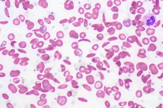
Schistocytes on a peripheral blood film
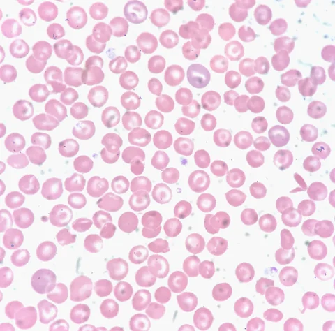
An individual ASH Image Bank image showing multiple schistocytes in a peripheral blood film.
ASH Image Bank · 61504 · Peripheral Blood with Multiple Schistocytes
MICA
A GUIDED SELECTION
Schistocytes, an Auer rod and sickle cells: three individual images for refining morphological description.
Describe the document or recording before looking at the explanation.
Use the teaching notes and original source to revisit your interpretation.
Return to the course, book or OSCE station associated with the resource.
TEACHING RESOURCE M0005
An individual ASH Image Bank image showing multiple schistocytes in a peripheral blood film.
ASH Image Bank · 61504 · Peripheral Blood with Multiple Schistocytes
TEACHING RESOURCE M0006
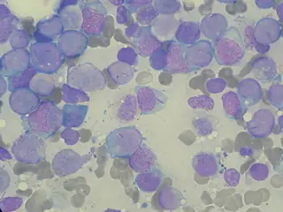
An ASH Image Bank photograph with an arrow identifying an Auer rod in a myeloid blast.
ASH Image Bank · 2523 · Auer Rod - 1.
TEACHING RESOURCE M0043
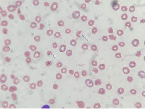
An ASH Image Bank image by Perla Vicari, with arrows indicating sickle cells.
ASH Image Bank · 63736 · Sickle cells (drepanocytes)
MICA IMAGES A0034
Elongated pointed erythrocytes suggesting an HbS haemoglobinopathy.
Ed Uthman from Houston, TX, USA — File : Sickle Cell Anemia (5610746554).jpg — CC BY 2.0 — https://commons.wikimedia.org/wiki/File%3ASickle_Cell_Anemia_(5610746554).jpg
ImagesMICA IMAGES A0035
Erythrocyte fragments identified and quantified on an appropriate film; haemolytic context is essential.
Moutaz Ghrewati; Anas Mahmoud; Tala Beilani; Karam Zakharia; Mehandar Kumar · How Should Complicated Cases of Thrombotic Thrombocytopenic Purpura With Positive Coombs Test Be Treated? · Cureus 2023;15(12):e50742, Figure 1 · CC BY 4.0 · https://assets.cureus.com/uploads/case_report/pdf/210009/20250210-74301-7pi8xp.pdf
MICA IMAGES A0036
Elongated cytoplasmic inclusions in myeloid cells; some presentations require urgent assessment.
Paulo Henrique Orlandi Mourao — File : Myeloblast with Auer rod smear 2010-01-27.JPG — CC BY-SA 3.0 — https://commons.wikimedia.org/wiki/File%3AMyeloblast_with_Auer_rod_smear_2010-01-27.JPG
MICA IMAGES A0044
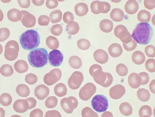
Lymphocyte population characterised morphologically, complemented by absolute count and immunophenotype.
Mary Ann Thompson (VashiDonsk at en.wikipedia) — File : Chronic lymphocytic leukemia.jpg — CC BY-SA 3.0 — https://commons.wikimedia.org/wiki/File%3AChronic_lymphocytic_leukemia.jpg
ImagesMICA IMAGES A0103
Recognise a target red cell without inferring a specific haemoglobinopathy.
Ed Uthman · Target Cells (51391831913).jpg · CC BY 2.0 · https://commons.wikimedia.org/wiki/File:Target_Cells_(51391831913).jpg
ImagesMICA IMAGES A0111
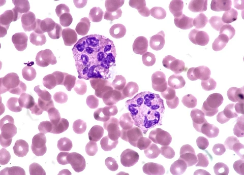
Recognise hypersegmentation without automatically attributing it to deficiency.
Ed Uthman — Hypersegmented_Neutrophils_(36831145373).jpg — CC BY 2.0
ImagesMICA IMAGES A0112
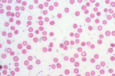
Describe dense round red cells, then distinguish mechanisms of haemolysis.
Ed Uthman — Spherocytes_(4670166434).jpg — CC BY 2.0