Flash information
- Observation
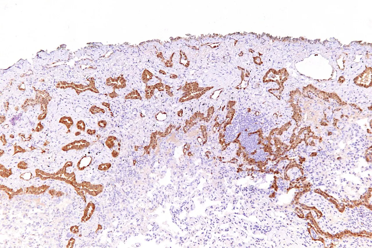
- The marker stains tumour structures.
- Limitation
- One positive marker cannot establish diagnosis.
Definition
Immunostaining in source-attributed mesothelioma.
Reading method
Locate brown staining and its cellular distribution.
Morphological description
The marker stains tumour structures.
Source context
The source context is epithelioid mesothelioma.
Differential diagnoses
Other tissues and tumours can express calretinin.
Clinical correlation
Integrate morphology and a marker panel.
Clinical impact
Oncological diagnosis requires specialist analysis.
Pitfalls and limitations
One positive marker cannot establish diagnosis.
Take-home message
Interpret a panel rather than colour alone.
Sources
Documentary review: 2026-10-14
Media source & rights
- Author
- Nephron
- Original source
- Nephron · Calrétinine : immunomarquage · CC BY-SA 3.0
- Licence
- CC BY-SA 3.0
- Changes
- Whole image retained; proportional resize and WebP conversion only. No clinical alteration. Derivative retains the original licence.
- Dimensions
- 1280 × 853 px
- Check
- 2026-10-14