Flash information
- Lesion
- Clinically suspected tophus
- Site
- Fingers and great toe
- Confirmation
- Crystals when diagnosis is uncertain
- Pitfall
- Coexisting infection is possible
Definition
A tophus is an organised tissue deposit of monosodium urate crystals. It reflects chronic crystal deposition.
Examination or reading method
Inspect hands, feet and other sites of nodules; assess consistency, mobility, pain and function. Look for skin breakdown.
Morphological description
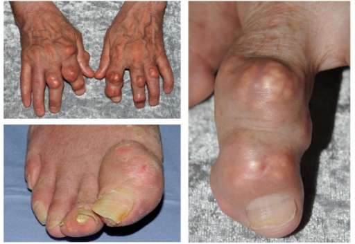
The source documents tophaceous gout. The panel shows finger and great-toe swellings, with pale areas beneath the skin.
Causes and diagnostic direction
Previous joint attacks and hyperuricaemia support the interpretation. Crystal identification in a sample provides confirmation when needed.
Differential diagnoses
Rheumatoid nodules, calcinosis and osteoarthritic nodes may enter the differential. Location, consistency and context matter more than colour alone.
Associated findings
Assess mobility, skin damage and renal or cardiovascular comorbidity. Establish treatment and flare history rather than inferring them from the photograph.
Red flags
A very painful joint with fever or a draining lesion requires assessment for infection, even in someone with established gout.
Pitfalls and limitations
Serum urate alone does not identify a nodule. The photograph does not establish current activity or adequacy of disease control.
Take-home message
Combine the deposit pattern, joint history and appropriate confirmation; consider infection when a lesion is inflamed.
Sources
- NICE NG219 — Gout: diagnosis and management (2022) ↗
- Arthritis Research UK Primary Care Centre, Keele University (E. Roddy) — document iconographique original / original image ↗
Documentary review: 2026-10-16
Media source & rights
- Author
- Arthritis Research UK Primary Care Centre, Keele University (E. Roddy)
- Original source
- Arthritis Research UK Primary Care Centre, Keele University (E. Roddy) — Tophaceous_gout.jpg — CC BY 2.0
- Licence
- CC BY 2.0
- Changes
- Main image converted to lossless WebP, EXIF orientation applied and metadata removed. Full field, colours and annotations retained; proportional thumbnails without upscaling or clinical retouch. Original media licence retained.
- Dimensions
- 512 × 353 px
- Check
- 2026-10-16