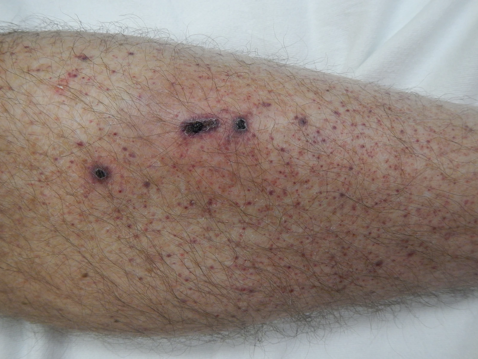
Flash information
- Size
- Pinpoint, usually ≤2 mm
- Elevation
- Flat macules
- Pressure
- Do not blanch
- Context
- Associated fever and bleeding
Definition
Petechiae are tiny purpuric lesions caused by superficial blood extravasation.
Examination or reading method
Confirm non-blanching and inspect the whole skin surface, conjunctivae and mouth.
Morphological description
Record clustered or diffuse distribution, recent onset, spread and accompanying bruises.
Causes and diagnostic direction
Causes include platelet disorders, infections and mechanically increased pressure after coughing or vomiting.
Differential diagnoses
Blanching lesions are not petechiae; any febrile haemorrhagic eruption requires clinical assessment.
Associated findings
Check for fever, headache, neck stiffness, confusion, cold extremities and rapid deterioration.
Red flags
Fever with rapidly spreading purpura or shock is an emergency; invasive infection is possible.
Pitfalls and limitations
Meningococcal rash may appear late: its absence cannot exclude severe infection.
Take-home message
Morphology identifies petechiae; clinical condition and evolution determine the priority of care.
Sources
- Royal Children’s Hospital Melbourne — Clinical Practice Guidelines: Petechiae and purpura · repères morphologiques ; recommandation pédiatrique ↗
- CDC — Meningococcal Disease Symptoms and Complications ↗
Documentary review: 2026-10-15
Media source & rights
- Author
- James Heilman, MD
- Original source
- James Heilman, MD — Petechia lower leg2.jpg — CC BY-SA 4.0 — https://commons.wikimedia.org/wiki/File:Petechia_lower_leg2.jpg
- Licence
- CC BY-SA 4.0
- Changes
- Orientation normalised and EXIF/GPS metadata removed. WebP conversion and proportional480/960/1600px derivatives, without upscaling, cropping or medical retouching. Derivatives shared under CC BY-SA4.0.
- Dimensions
- 4608 × 3456 px
- Check
- 2026-10-15