Flash information
- Technique
- Upper GI endoscopy
- Observation
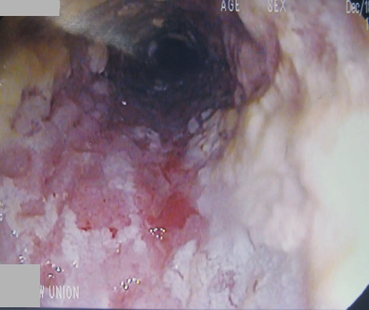
- Whitish deposits cover inflamed mucosa.
- Limitation
- The photograph cannot identify the fungal species.
Definition
Whitish oesophageal plaques identified as candidiasis.
Reading method
Assess distribution and underlying mucosa.
Morphological description
Whitish deposits cover inflamed mucosa.
Interpretation and diagnostic limits
The source diagnosis guides image reading.
Differential diagnoses
Compare deposits, inflammatory lesions and other infections.
Clinical correlation
Ask about dysphagia, odynophagia and risk factors.
Clinical impact
Marked dysphagia warrants assessment.
Pitfalls
The photograph cannot identify the fungal species.
Take-home message
Integrate endoscopic appearance and context.
Media source & rights
- Author
- James Heilman, MD
- Original source
- James Heilman, MD · Plaques œsophagiennes — candidose attribuée par la source · CC BY-SA 3.0
- Licence
- CC BY-SA 3.0
- Changes
- Full image retained; WebP conversion and proportional resize. No clinical alteration. Derivative retains the source licence.
- Dimensions
- 1280 × 1078 px
- Check
- 2026-10-14