Flash information
- Lesion
- Tissue defect
- Description
- Site, size, base, edges
- Common causes
- H. pylori and NSAIDs
- Complication
- Bleeding or perforation
Definition
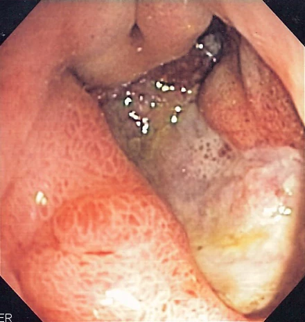
A gastric ulcer is a tissue defect deeper than an erosion.
Examination or reading method
Identify the site, obtain a clear view and document size, edges, base and bleeding signs.
Morphological description
Describe crater, fibrin, converging folds and any visible vessel; one photograph may not permit complete classification.
Causes and diagnostic direction
H. pylori and NSAIDs are common causes; also assess for malignant lesions.
Differential diagnoses
Distinguish erosion from ulcer; visual appearance alone does not always separate benign ulceration from malignancy.
Associated findings
Assess pain, anaemia, bleeding and predisposing medicines; H. pylori diagnosis requires appropriate testing.
Red flags
Haematemesis, melaena, faintness or sudden abdominal pain suggest complications requiring urgent assessment.
Pitfalls and limitations
A fibrinous base does not establish benignity; endoscopy and tissue sampling form part of medical assessment.
Take-home message
Describe the ulcer, assess complications and determine its cause; do not infer benignity from an image.
Sources
- Gastric Ulcer - StatPearls - NCBI Bookshelf - NIH ↗
- Peptic Ulcers (Stomach or Duodenal Ulcers) - NIDDK ↗
Documentary review: 2026-10-12
Media source & rights
- Author
- Samir
- Original source
- Samir · Ulcère gastrique profond — image endoscopique · https://commons.wikimedia.org/wiki/File:Deep_gastric_ulcer.png · CC BY-SA 3.0
- Licence
- CC BY-SA 3.0
- Changes
- EXIF omitted; aspect ratio preserved; proportional resize and WebP conversion only. No medical pixels retouched.
- Dimensions
- 440 × 464 px
- Check
- 2026-10-11