Flash information
- Observation
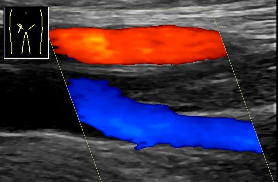
- Red and blue depend on flow direction relative to the probe.
- Limitation
- Colour alone cannot prove thrombosis.
Definition
Image acquired in source-documented femoral DVT.
Reading method
Identify vessels and areas of colour signal.
Morphological description
Red and blue depend on flow direction relative to the probe.
Source context
Thrombosis attribution comes from the source.
Differential diagnoses
Distinguish flow abnormalities from non-compressibility.
Clinical correlation
Integrate compression, symptoms and clinical probability.
Clinical impact
Associated dyspnoea may warrant assessment for pulmonary embolism.
Pitfalls and limitations
Colour alone cannot prove thrombosis.
Take-home message
Read the complete examination, not colour alone.
Sources
Documentary review: 2026-10-14
Media source & rights
- Author
- Mikael Häggström
- Original source
- Mikael Häggström · Échographie de TVP fémorale · CC0 1.0
- Licence
- CC0 1.0
- Changes
- Whole image retained; proportional resize and WebP conversion only. No clinical alteration. Derivative retains the original licence.
- Dimensions
- 960 × 630 px
- Check
- 2026-10-14