Flash information
- Observation
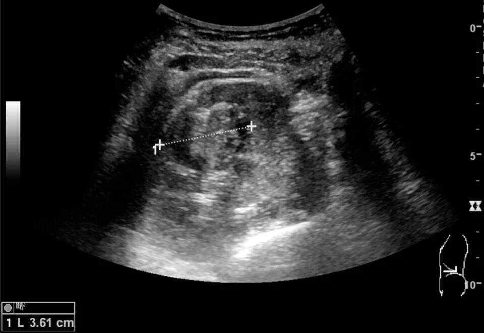
- Source markers measure the solid lesion.
- Limitation
- Ultrasound alone is not histological proof.
Definition
A solid cortical renal mass on ultrasound.
Reading method
Compare mass, cortex and renal sinus.
Morphological description
Source markers measure the solid lesion.
Source context
The source reports later carcinoma confirmation.
Differential diagnoses
Differential assessment requires additional imaging.
Clinical correlation
Correlate characterisation and diagnostic confirmation.
Clinical impact
One image cannot determine prognosis.
Pitfalls and limitations
Ultrasound alone is not histological proof.
Take-home message
Separate visible mass from confirmed diagnosis.
Sources
Documentary review: 2026-10-14
Media source & rights
- Author
- Kristoffer Lindskov Hansen, Michael Bachmann Nielsen and Caroline Ewertsen
- Original source
- Kristoffer Lindskov Hansen, Michael Bachmann Nielsen and Caroline Ewertsen · Masse rénale solide : échographie · CC BY 4.0
- Licence
- CC BY 4.0
- Changes
- Whole image retained; proportional resize and WebP conversion only. No clinical alteration. Derivative retains the original licence.
- Dimensions
- 960 × 662 px
- Check
- 2026-10-14