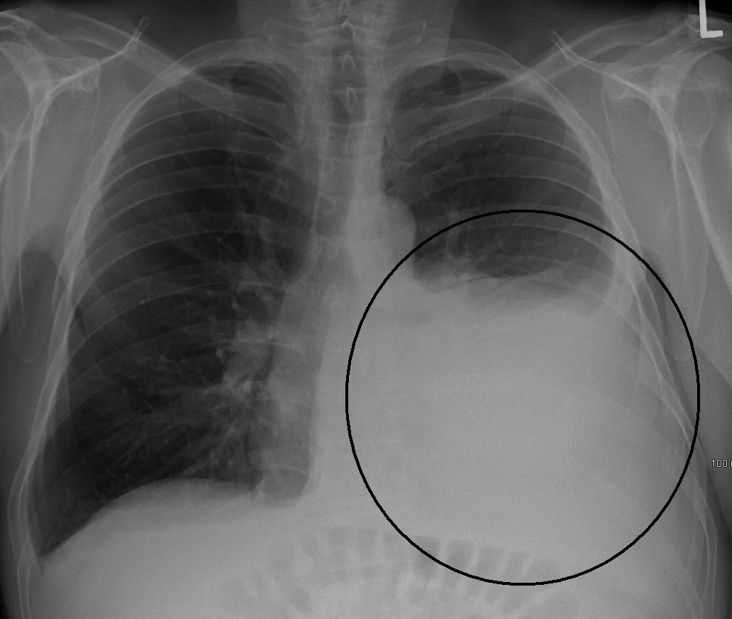
Flash information
- Compartment
- Pleural space
- Finding
- Costophrenic blunting
- Contour
- Possible meniscus
- Complement
- Thoracic ultrasound
Definition
Pleural effusion is abnormal fluid accumulation between the pleural layers.
Examination or reading method
Check upright or supine position, compare both bases and assess a meniscus or dependent opacity.
Morphological description
Describe side, apparent size, consequences and possible loculation; opacity alone does not determine cause.
Causes and diagnostic direction
Heart failure, infection, malignancy, pulmonary embolism and systemic inflammatory disease are possible directions.
Differential diagnoses
Differentiate fluid, pleural thickening and pulmonary opacity; ultrasound better identifies fluid.
Associated findings
Ultrasound assesses mobility, contents and septa and identifies the diaphragm to distinguish effusion from ascites.
Red flags
Respiratory distress or infection changes urgency; ultrasound features can guide appropriate investigation.
Pitfalls and limitations
Anechoic fluid does not define a transudate; supine radiography may conceal substantial fluid.
Take-home message
Locate the fluid, assess clinical consequences and investigate its cause with targeted testing.
Sources
- Pleural Effusion - StatPearls - NCBI Bookshelf ↗
- Thoracic and Lung Ultrasound - StatPearls - NCBI Bookshelf ↗
Documentary review: 2026-10-12
Media source & rights
- Author
- James Heilman, MD
- Original source
- James Heilman, MD · Radiographie : épanchement pleural gauche · https://commons.wikimedia.org/wiki/File:Effusionhalf.PNG · CC BY-SA 3.0
- Licence
- CC BY-SA 3.0
- Changes
- EXIF omitted; aspect ratio preserved; proportional resize and WebP conversion only. No medical pixels retouched.
- Dimensions
- 1030 × 871 px
- Check
- 2026-10-11