Flash information
- Line
- Visceral pleural edge
- Peripheral region
- Absent vascular markings
- Projection
- Upright or supine
- Urgency
- Assess clinical stability
Definition
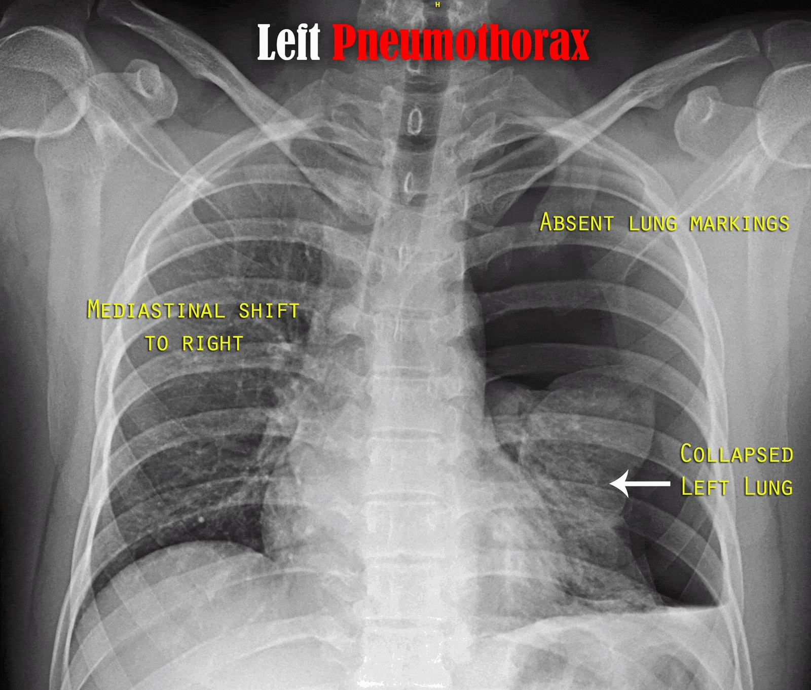
Pneumothorax is air within the pleural space, separating lung from chest wall.
Examination or reading method
Check projection and position; identify the pleural line and absent vessels outside it.
Morphological description
Describe side, visible extent and lung status; supine air may produce an unusually deep costophrenic sulcus.
Causes and diagnostic direction
Contexts include trauma, medical procedures or spontaneous pneumothorax with or without underlying lung disease.
Differential diagnoses
Skin folds, bullae and scapular edges may mimic a pleural line; check for markings beyond it.
Associated findings
Assess mediastinal shift and diaphragmatic depression alongside vital signs and symptoms.
Red flags
Tension pneumothorax is a clinical emergency; compatible instability must not await radiological confirmation.
Pitfalls and limitations
Supine radiography may underestimate or miss pneumothorax; mediastinal shift alone does not establish tension.
Take-home message
Identify pleural air and immediately assess its clinical consequences.
Sources
- Spontaneous Pneumothorax - StatPearls - NCBI Bookshelf ↗
- Tension Pneumothorax - StatPearls - NCBI Bookshelf ↗
Documentary review: 2026-10-15
Media source & rights
- Author
- Karthik Easvur
- Original source
- Karthik Easvur, X-ray_of_pneumothorax_signs.jpg, CC BY-SA 3.0, Wikimedia Commons; conversion WebP et réduction proportionnelle.
- Licence
- CC BY-SA 3.0
- Changes
- Full original field and annotations retained; WebP conversion, EXIF/GPS metadata removed, proportional derivatives without upscaling, cropping or medical retouching. Derivatives shared under the same CC BY-SA 3.0 licence.
- Dimensions
- 1980 × 1684 px
- Check
- 2026-10-15