Flash information
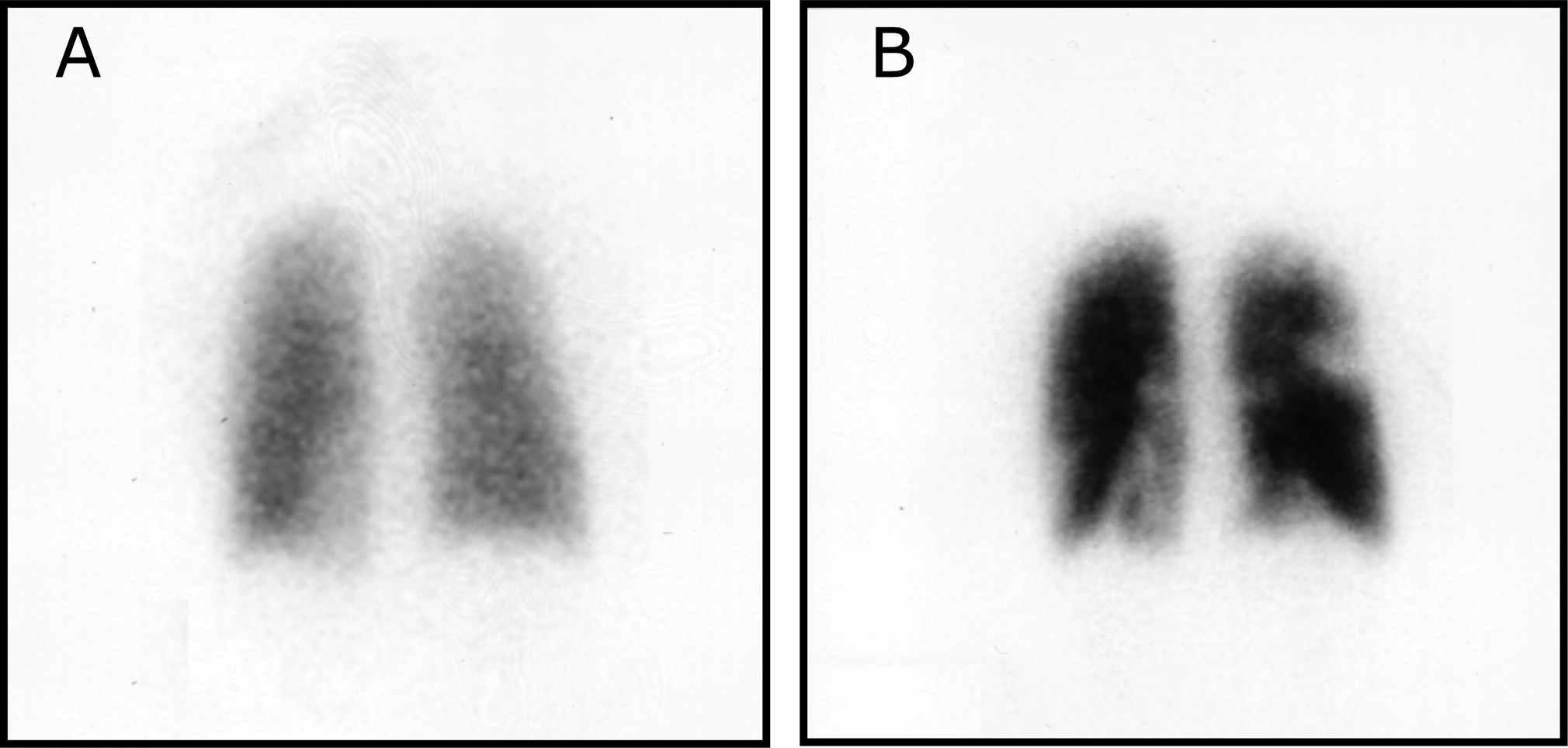
- Panel A
- Ventilation
- Panel B
- Perfusion
- Principle
- Compare matching regions
- Technique
- Posterior planar views
Definition
Mismatch means reduced perfusion in a region with relatively preserved ventilation; its distribution matters for interpretation.
Examination or reading method
Check orientation, technique and quality; compare the same regions on ventilation and perfusion using all available projections.
Morphological description
A shows relatively uniform ventilation; B contains several regional defects. This pair represents only part of the complete examination.
Causes and diagnostic direction
Segmental vascular mismatch suggests embolic obstruction. The source reports a patient with already documented pulmonary embolism.
Differential diagnoses
Artefacts, lung disease and vascular abnormalities may complicate interpretation. Matched ventilation–perfusion defects have a different meaning.
Associated findings
Integrate clinical probability, symptoms, risk factors and chest imaging; abnormalities on an isolated view do not establish the diagnosis alone.
Red flags
Shock, hypotension or respiratory distress requires an urgent pathway adapted to stability; do not await routine scintigraphy.
Pitfalls and limitations
Do not automatically apply validated SPECT thresholds to these planar views. Scintigraphy does not directly show the thrombus.
Take-home message
Compare ventilation and perfusion, verify vascular distribution and review the complete study; distinguish learning the sign from treatment decisions.
Sources
- Bajc et al. EANM guideline for ventilation/perfusion SPECT for diagnosis of pulmonary embolism and beyond (2019) ↗
- Westgate and FitzGerald. Pulmonary Embolism in a Woman Taking Oral Contraceptives and Valdecoxib (2005), Figure 1 ↗
Documentary review: 2026-10-15
Media source & rights
- Author
- Elizabeth J. Westgate; Garret A. FitzGerald
- Original source
- Elizabeth J. Westgate; Garret A. FitzGerald — Pulmonary Embolism in a Woman Taking Oral Contraceptives and Valdecoxib (2005), Figure1 — CC BY2.5 — https://journals.plos.org/plosmedicine/article?id=10.1371/journal.pmed.0020197
- Licence
- CC BY 2.5
- Changes
- Full original field and annotations retained; WebP conversion, EXIF/GPS metadata removed, proportional derivatives without upscaling, cropping or medical retouching.
- Dimensions
- 2020 × 964 px
- Check
- 2026-10-15