Flash information
- Sign
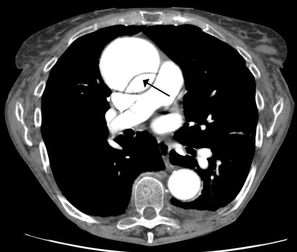
- Intimal flap
- Site
- Ascending aorta
- Classification
- Type A when ascending involvement is confirmed
- Priority
- Urgent aortic specialist assessment
Definition
Dissection separates layers of the aortic wall, creating channels divided by a flap. Ascending involvement defines Stanford type A.
Examination or reading method
Follow the aorta throughout the study and reconstructions. Assess entry tear, extent, branch involvement and complications; check motion artefacts.
Morphological description
A thin linear structure crosses the enhanced ascending aortic lumen at the arrow tip. The source identifies ascending aortic dissection.
Causes and diagnostic direction
Integrate acute pain, vascular examination and aortic history. Hypertension or known aortopathy may guide interpretation but cannot be inferred from the image.
Differential diagnoses
Pulsation artefact may mimic a flap. Intramural haematoma and penetrating aortic ulcer are other acute aortic syndromes with distinct criteria.
Associated findings
Assess pulse asymmetry, neurological deficit and visceral malperfusion. Evaluate the aortic valve and pericardium with appropriate investigations.
Red flags
Acute type A dissection requires immediate surgical assessment. Shock, tamponade and malperfusion are major complications to investigate.
Pitfalls and limitations
Do not label true and false lumina from their apparent size on this slice alone. Absence of a complication in this field does not exclude it.
Take-home message
Identify the flap, establish ascending involvement and raise the alarm without delaying for a complete teaching exercise.
Sources
- ACC/AHA — Aortic disease guideline: key perspectives, part 2 (2022) ↗
- James Heilman, MD — document iconographique original / original image ↗
Documentary review: 2026-10-16
Media source & rights
- Author
- James Heilman, MD
- Original source
- James Heilman, MD — DissectionCT.png — CC BY-SA 3.0
- Licence
- CC BY-SA 3.0
- Changes
- Main image converted to lossless WebP, EXIF orientation applied and metadata removed. Full field, colours and annotations retained; proportional thumbnails without upscaling or clinical retouch. Original media licence retained.
- Dimensions
- 1030 × 875 px
- Check
- 2026-10-16