Flash information
- Sign
- Filling defect
- Examination
- CT pulmonary angiography
- Severity
- Haemodynamics and right ventricle
- Pitfall
- Central location alone does not grade severity
Definition
Thrombotic pulmonary embolism obstructs part of the pulmonary arterial bed. On CT angiography, thrombus appears as a defect within contrast-enhanced blood.
Examination or reading method
Check enhancement and artefacts before following arteries through consecutive slices. Compare reconstructions with the full series.
Morphological description
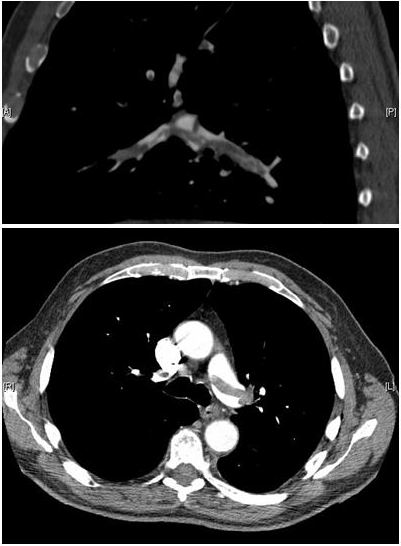
Both views show lower-attenuation areas within enhanced arteries. The original case describes a saddle embolus with bilateral extension.
Causes and diagnostic direction
Relate the radiological diagnosis to clinical probability and thromboembolic context. This image alone establishes neither clot origin nor age.
Differential diagnoses
Contrast mixing, respiratory motion and partial-volume effects can mimic a defect. Multiplanar review of consecutive images is necessary.
Associated findings
Document blood pressure, oxygenation and right ventricular dysfunction; biomarkers and clinical scores complement risk stratification.
Red flags
Persistent hypotension, shock or cardiopulmonary deterioration requires urgent care. Reperfusion decisions depend on specialist risk assessment.
Pitfalls and limitations
A saddle embolus alone does not classify severity. These two views cannot reliably measure ventricular ratio or exclude other emboli.
Take-home message
Recognise the filling defect, confirm it on the full study, then assess clinical tolerance and the right heart.
Sources
- AHA/ACC — Acute pulmonary embolism guideline: key points (2026) ↗
- Myat & Ahsan — Pulmonary embolism: original image and case report (2007) ↗
- Aung Myat et Arif Ahsan — document iconographique original / original image ↗
Documentary review: 2026-10-16
Media source & rights
- Author
- Aung Myat et Arif Ahsan
- Original source
- Aung Myat et Arif Ahsan — Pulmonary_embolism_CTPA.JPEG — CC BY 2.0
- Licence
- CC BY 2.0
- Changes
- Main image converted to lossless WebP, EXIF orientation applied and metadata removed. Full field, colours and annotations retained; proportional thumbnails without upscaling or clinical retouch. Original media licence retained.
- Dimensions
- 401 × 546 px
- Check
- 2026-10-16