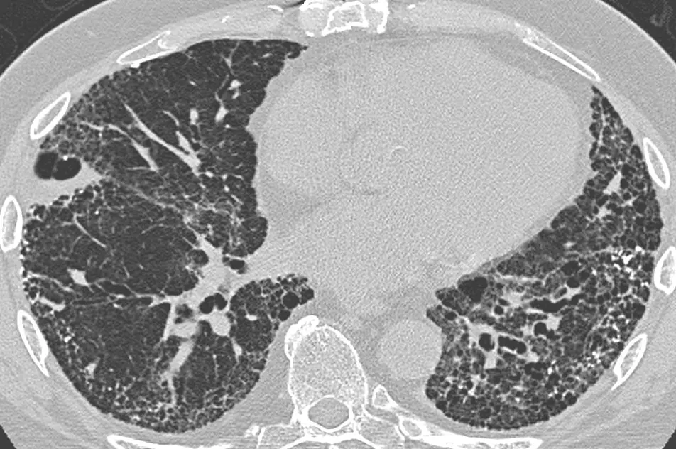
Flash information
- Morphology
- Clustered visible-walled cysts
- Site
- Often subpleural
- Association
- Distortion and fibrosis
- Aetiology
- Determined in context
Definition
Honeycombing describes clustered cystic spaces within a lung remodelled by fibrosis.
Examination or reading method
Follow spaces on contiguous sections and reconstructions; assess their relationship to pleura and bronchi.
Morphological description
Describe clustering, possible layering, basal or other predominance and accompanying volume loss.
Causes and diagnostic direction
Usual interstitial pneumonia may show this appearance; other fibrosing interstitial diseases can also produce it.
Differential diagnoses
Paraseptal emphysema and traction bronchiectasis can resemble honeycombing; review the complete series.
Associated findings
Assess reticulation, traction bronchiectasis and distribution; review exposure, connective-tissue disease and lung function.
Red flags
Fibrotic extent and evolution contribute to prognosis; symptoms and lung function complete the assessment.
Pitfalls and limitations
A usual interstitial pneumonia pattern does not automatically establish idiopathic pulmonary fibrosis.
Take-home message
Recognise cystic remodelling, then determine the pattern and cause through multidisciplinary correlation.
Reticulation · CT
Reticulation consists of intersecting linear opacities producing a network-like appearance. Describe fine or coarse appearance, subpleural or diffuse distribution and associated volume loss.
Sources
- Idiopathic Interstitial Pneumonias - PMC ↗
- Idiopathic Pulmonary Fibrosis (an Update) and Progressive Pulmonary Fibrosis in Adults: An Official ATS/ERS/JRS/ALAT Clinical Practice Guideline - PMC ↗
- Plain Film and HRCT Diagnosis of Interstitial Lung Disease - Diseases of the Chest, Breast, Heart and Vessels 2019-2022 - NCBI Bookshelf ↗
- Fleischner Society: Glossary of Terms for Thoracic Imaging ↗
Documentary review: 2026-10-12
Media source & rights
- Author
- Yale Rosen from USA
- Original source
- Yale Rosen from USA, CT_scan_in_usual_interstitial_pneumonia_(UIP).jpg, CC BY-SA 2.0, Wikimedia Commons; conversion WebP et réduction proportionnelle.
- Licence
- CC BY-SA 2.0
- Changes
- Conversion WebP, redimensionnement proportionnel sans recadrage ni annotations ajoutées; EXIF retiré. Dérivé écran proportionnel ≤1280 px, WebP qualité76 ; vignette ≤320 px qualité72. Source acquise et dérivé initial conservés sans modification ; aucune retouche médicale, aucun recadrage.
- Dimensions
- 952 × 632 px
- Check
- 2026-10-11T20:52:42.061125+00:00