Original MICA image-reading exercise, unscored. No clinical narrative absent from the source is added.
- Describe visible findings before proposing an interpretation.
- Suggest an interpretation and at least one image limitation.
- Which clinical findings or investigations would complement this observation?
Reveal the explained reading
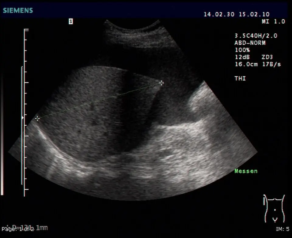
Ascites on ultrasound
- Technique
- Ultrasound
- Observation
- Anechoic fluid abuts abdominal structures.
- Limitation
- Black appearance does not identify fluid composition.
Morphological description
Anechoic fluid abuts abdominal structures.
Interpretation and diagnostic limits
The source documents ascites without establishing its cause here.
Differential diagnoses
Distinguish free fluid, loculated collections and cystic structures.
Pitfalls
Black appearance does not identify fluid composition.
Take-home message
Recognise fluid before investigating its cause.
Media source & rights
- Author
- Drahreg01
- Original source
- Drahreg01 · Ascite à l’échographie · CC BY-SA 3.0
- Licence
- CC BY-SA 3.0
- Changes
- Full image retained; WebP conversion and proportional resize. No clinical alteration. Derivative retains the source licence.
- Dimensions
- 960 × 783 px
- Check
- 2026-10-14