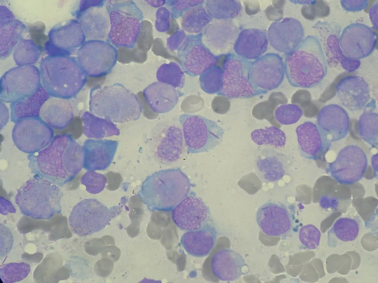
Original MICA image-reading exercise, unscored. No clinical narrative absent from the source is added.
- Describe visible findings before proposing an interpretation.
- Suggest an interpretation and at least one image limitation.
- Which clinical findings or investigations would complement this observation?
Reveal the explained reading
Auer rods · cytology
- Location
- Cytoplasm
- Appearance
- Elongated azurophilic inclusion
- Direction
- Myeloid differentiation
- Warning
- Suspected promyelocytic leukaemia
Morphological description
Rods may be isolated or bundled; describe their distribution and the cell morphology.
Causes and diagnostic direction
They suggest myeloid proliferation; bundles within promyelocytes raise acute promyelocytic leukaemia.
Differential diagnoses
Distinguish morphology from artefacts; classification also requires immunophenotyping and genetics.
Pitfalls and limitations
One Auer rod does not establish promyelocytic leukaemia; absent rods do not exclude it.
Take-home message
Recognise the inclusion, escalate according to context and confirm classification with required investigations.
Sources
- Acute Promyelocytic Leukemia - StatPearls - NCBI Bookshelf ↗
- Acute promyelocytic leukemia: preventing early complications and late toxicities - PMC ↗
Documentary review: 2026-10-12
Media source & rights
- Author
- Paulo Henrique Orlandi Mourao
- Original source
- Paulo Henrique Orlandi Mourao — File : Myeloblast with Auer rod smear 2010-01-27.JPG — CC BY-SA 3.0 — https://commons.wikimedia.org/wiki/File%3AMyeloblast_with_Auer_rod_smear_2010-01-27.JPG
- Licence
- CC BY-SA 3.0
- Changes
- EXIF supprimé ; redimensionnement proportionnel ; conversion WebP ; aucun recadrage ni retouche médicale. Dérivé écran proportionnel ≤1280 px, WebP qualité76 ; vignette ≤320 px qualité72. Source acquise et dérivé initial conservés sans modification ; aucune retouche médicale, aucun recadrage.
- Dimensions
- 1280 × 960 px
- Check
- 2026-10-11T20:56:14.343068+00:00