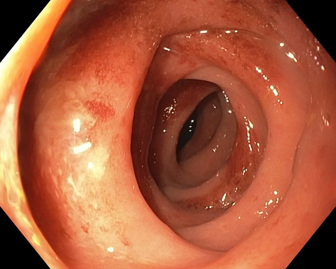
Original MICA image-reading exercise, unscored. No clinical narrative absent from the source is added.
- Describe visible findings before proposing an interpretation.
- Suggest an interpretation and at least one image limitation.
- Which clinical findings or investigations would complement this observation?
Reveal the explained reading
Segmental colitis associated with diverticulosis
- Technique
- Colonoscopy
- Observation
- Erythema alters the vascular pattern.
- Limitation
- One image cannot demonstrate rectal sparing.
Morphological description
Erythema alters the vascular pattern.
Interpretation and diagnostic limits
The source identifies diverticulosis-associated colitis.
Differential diagnoses
Consider inflammatory bowel disease, infection and ischaemia.
Pitfalls
One image cannot demonstrate rectal sparing.
Take-home message
Distribution matters as much as appearance.
Sources
Documentary review: 2026-10-14
Media source & rights
- Author
- Samir
- Original source
- Samir · Colite segmentaire associée à une diverticulose · CC BY-SA 4.0
- Licence
- CC BY-SA 4.0
- Changes
- Full image retained; WebP conversion and proportional resize. No clinical alteration. Derivative retains the source licence.
- Dimensions
- 1280 × 1024 px
- Check
- 2026-10-14