Original MICA image-reading exercise, unscored. No clinical narrative absent from the source is added.
- Describe visible findings before proposing an interpretation.
- Suggest an interpretation and at least one image limitation.
- Which clinical findings or investigations would complement this observation?
Reveal the explained reading
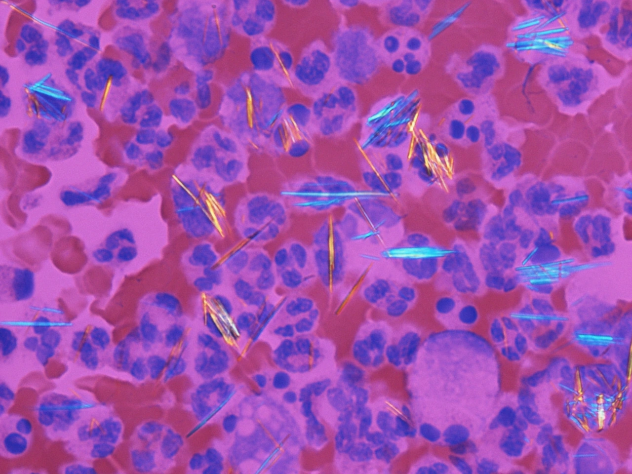
Urate crystals: polarised light
- Shape
- Needles
- Technique
- Compensated polarised light
- Reading
- Check orientation
- Pitfall
- Crystals do not exclude infection
Morphological description
The field shows numerous long pointed yellow or blue structures among inflammatory cells. Some needles overlap cells.
Causes and diagnostic direction
The source identifies urate crystals. In practice, needle shape and negative birefringence support identification after correct technical assessment.
Differential diagnoses
Calcium pyrophosphate crystals are often rhomboid or rod-shaped and weakly positively birefringent. Debris and artefacts must also be distinguished.
Pitfalls and limitations
Colour alone cannot establish the sign of birefringence without the compensator axis. A still field cannot replace examination of the sample.
Take-home message
Describe the needles, verify polarised-light findings and integrate the clinical picture; never use crystals alone to exclude infection.
Sources
- NICE NG219 — Gout: diagnosis and management (2022) ↗
- Gabriel Caponetti — document iconographique original / original image ↗
Documentary review: 2026-10-16
Media source & rights
- Author
- Gabriel Caponetti
- Original source
- Gabriel Caponetti — Gout_-_monosodium_urate_crystals_(20X,_polarized,_red_compensator).jpg — CC BY-SA 3.0
- Licence
- CC BY-SA 3.0
- Changes
- Main image converted to lossless WebP, EXIF orientation applied and metadata removed. Full field, colours and annotations retained; proportional thumbnails without upscaling or clinical retouch. Original media licence retained.
- Dimensions
- 1280 × 960 px
- Check
- 2026-10-16