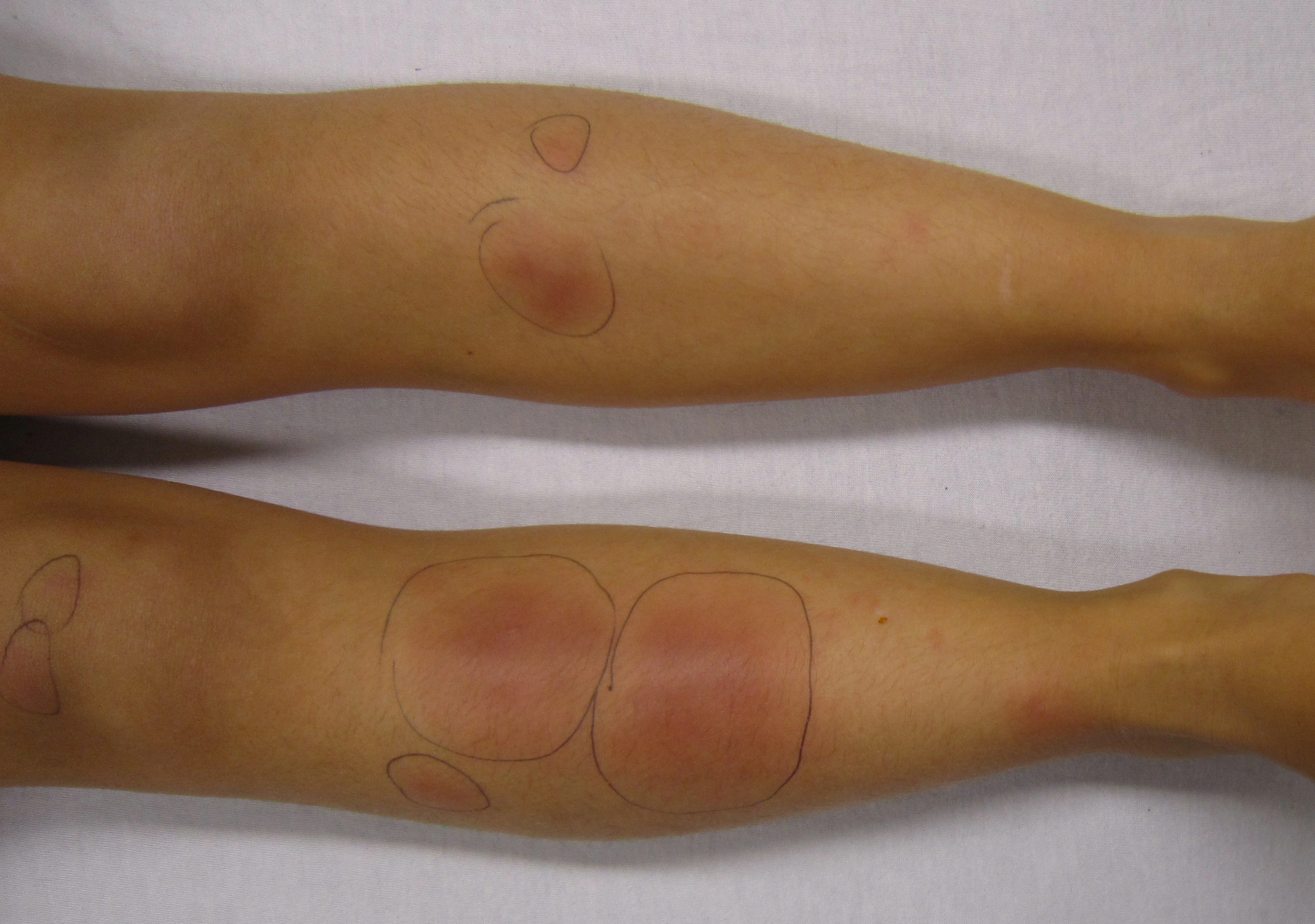
Original MICA image-reading exercise, unscored. No clinical narrative absent from the source is added.
- Describe visible findings before proposing an interpretation.
- Suggest an interpretation and at least one image limitation.
- Which clinical findings or investigations would complement this observation?
Reveal the explained reading
Erythema nodosum: nodules on the legs
- Appearance
- Inflammatory nodules
- Site
- Anterior shins
- Examination
- Palpation is essential
- Next step
- Investigate the underlying context
Morphological description
The photograph shows several rounded erythematous areas of different sizes on both legs. Ink circles are landmarks added by the source.
Causes and diagnostic direction
History should explore recent infection, medication, bowel symptoms and sarcoidosis. Investigations depend on associated findings and local epidemiology, including tuberculosis.
Differential diagnoses
Superficial thrombophlebitis, skin infection and other panniculitides may cause painful lesions. Ulceration or an atypical location should prompt reconsideration.
Pitfalls and limitations
Redness alone proves neither panniculitis nor its cause. Warmth, pain and subcutaneous depth cannot be assessed from this image.
Take-home message
First describe location and distribution; confirm nodules by palpation before directing the investigation of their cause.
Sources
Documentary review: 2026-10-16
Media source & rights
- Author
- James Heilman, MD
- Original source
- James Heilman, MD — ENlegs.JPG — CC BY-SA 3.0
- Licence
- CC BY-SA 3.0
- Changes
- Main image converted to lossless WebP, EXIF orientation applied and metadata removed. Full field, colours and annotations retained; proportional thumbnails without upscaling or clinical retouch. Original media licence retained.
- Dimensions
- 3074 × 2160 px
- Check
- 2026-10-16