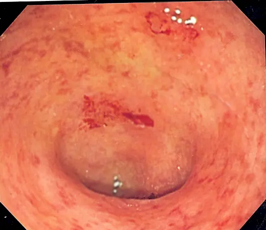
Original MICA image-reading exercise, unscored. No clinical narrative absent from the source is added.
- Describe visible findings before proposing an interpretation.
- Suggest an interpretation and at least one image limitation.
- Which clinical findings or investigations would complement this observation?
Reveal the explained reading
Inflamed colonic mucosa · endoscopy
- Surface
- Granularity
- Vessels
- Reduced markings
- Fragility
- Friability or bleeding
- Distribution
- Continuous or segmental
Morphological description
Record granularity, loss of markings, friability, erosions, ulcers and spontaneous bleeding; findings may coexist.
Causes and diagnostic direction
Inflammatory bowel disease, infection, ischaemia and medicines can cause colitis.
Differential diagnoses
Continuous involvement from the rectum suggests ulcerative colitis and must be distinguished from other colitides.
Pitfalls and limitations
Granularity alone does not diagnose ulcerative colitis; treatment and preparation quality alter appearance.
Take-home message
Describe inflammation and distribution, then correlate infectious, clinical and pathological evidence.
Sources
Documentary review: 2026-10-12
Media source & rights
- Author
- Samir
- Original source
- Samir — File : UC granularity.png — CC BY-SA 3.0 — https://commons.wikimedia.org/wiki/File%3AUC_granularity.png
- Licence
- CC BY-SA 3.0
- Changes
- EXIF supprimé ; redimensionnement proportionnel ; conversion WebP ; aucun recadrage ni retouche médicale. Dérivé écran proportionnel ≤1280 px, WebP qualité76 ; vignette ≤320 px qualité72. Source acquise et dérivé initial conservés sans modification ; aucune retouche médicale, aucun recadrage.
- Dimensions
- 544 × 471 px
- Check
- 2026-10-11T20:53:34.493315+00:00