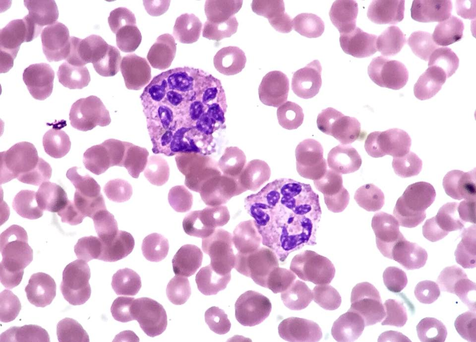
Original MICA image-reading exercise, unscored. No clinical narrative absent from the source is added.
- Describe visible findings before proposing an interpretation.
- Suggest an interpretation and at least one image limitation.
- Which clinical findings or investigations would complement this observation?
Reveal the explained reading
Hypersegmented neutrophils: blood film
- Sign
- Nuclear hypersegmentation
- Document
- Authentic blood film
- Correlate with
- CBC, vitamins, medication
- Pitfall
- Non-specific finding
Morphological description
Both visible purple cells have nuclei divided into numerous lobes. The field also contains red cells with varying size and shape.
Causes and diagnostic direction
Vitamin B12 or folate deficiency is one possibility; medication can also affect maturation. Here, the author reports chemotherapy.
Differential diagnoses
Do not confuse connected lobes of one nucleus with overlapping cells. Dysplasia requires assessment of the whole film and clinical context.
Pitfalls and limitations
The film alone cannot diagnose autoimmune gastritis. Absence of macrocytosis or anaemia also does not exclude B12 deficiency.
Take-home message
Name hypersegmentation, then evaluate possible causes; respect the source context rather than assigning an undocumented aetiology.
Sources
- NICE NG239 — Vitamin B12 deficiency (2024) ↗
- Ed Uthman — document iconographique original / original image ↗
Documentary review: 2026-10-16
Media source & rights
- Author
- Ed Uthman
- Original source
- Ed Uthman — Hypersegmented_Neutrophils_(36831145373).jpg — CC BY 2.0
- Licence
- CC BY 2.0
- Changes
- Main image converted to lossless WebP, EXIF orientation applied and metadata removed. Full field, colours and annotations retained; proportional thumbnails without upscaling or clinical retouch. Original media licence retained.
- Dimensions
- 960 × 690 px
- Check
- 2026-10-16