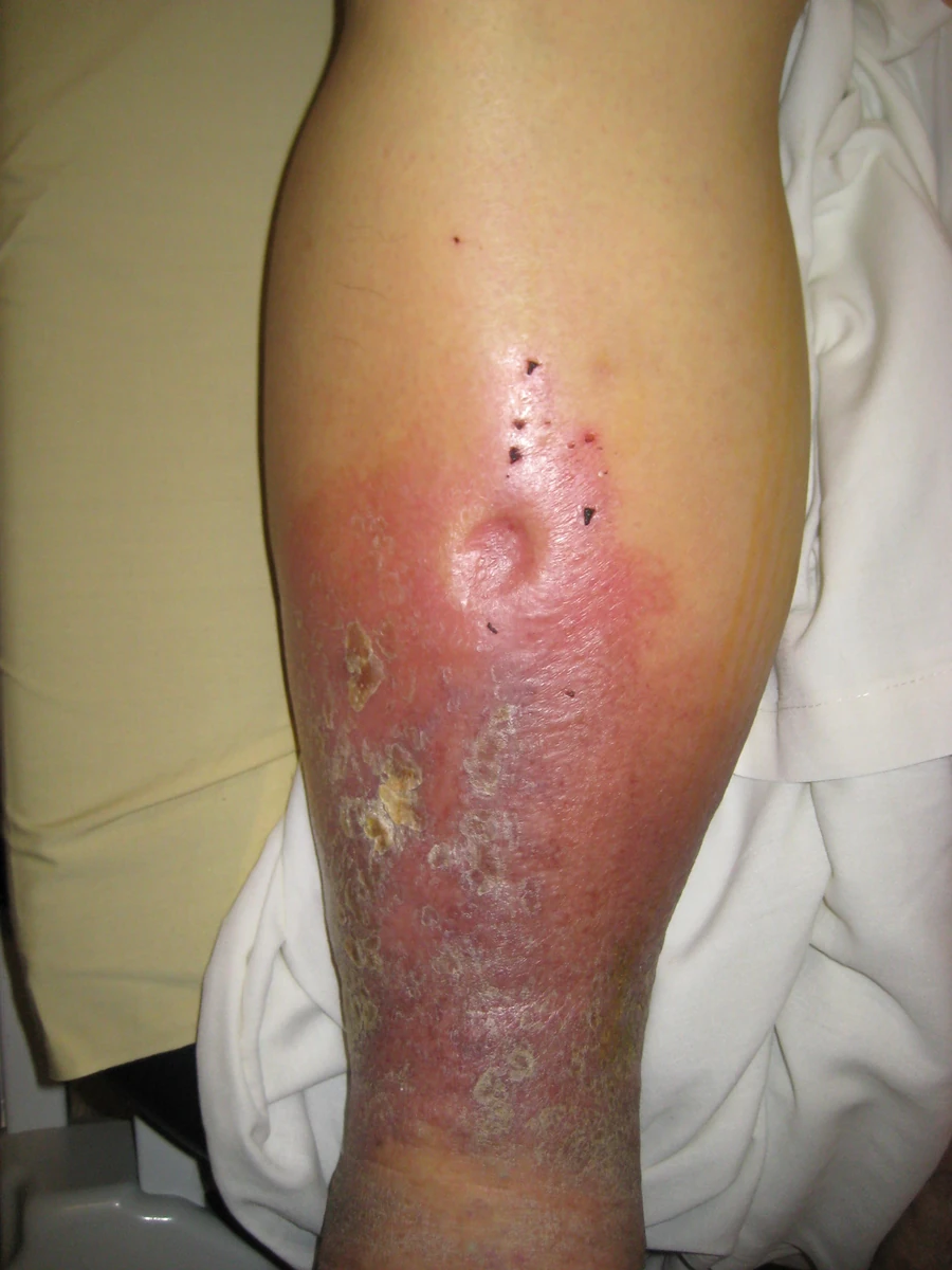
Original MICA image-reading exercise, unscored. No clinical narrative absent from the source is added.
- Describe visible findings before proposing an interpretation.
- Suggest an interpretation and at least one image limitation.
- Which clinical findings or investigations would complement this observation?
Reveal the explained reading
Pitting oedema
- Sign
- Persistent indentation
- Mechanisms
- Pressure, albumin, permeability, lymph
- Distribution
- Unilateral or bilateral
- Urgency
- Dyspnoea or acutely painful limb
Morphological description
Describe location, extent, symmetry, pain, colour, temperature and duration. Check sacral oedema in a bedbound patient.
Causes and diagnostic direction
Mechanisms include raised hydrostatic pressure, reduced oncotic pressure, increased permeability or impaired lymphatic drainage; several may coexist.
Differential diagnoses
Lymphoedema may pit initially and later become firm. Lipoedema often spares the feet; myxoedema and a mass are different findings.
Pitfalls and limitations
Pitting is a sign, not a diagnosis of heart failure. A photograph establishes neither cause, intravascular volume nor severity.
Take-home message
Confirm pitting, describe distribution and investigate the cause. New unilateral leg swelling and acute dyspnoea immediately change clinical priority.
Sources
- Edema - Clinical Methods - NCBI Bookshelf ↗
- Pitting Edema Causes, Grading & Treatment ↗
- Peripheral Edema - StatPearls - NCBI Bookshelf ↗
- Diagnostic approach to lower limb edema - PMC - NIH ↗
- Peripheral Edema: Evaluation and Management in Primary ... ↗
Documentary review: 2026-10-12
Media source & rights
- Author
- James Heilman, MD
- Original source
- James Heilman, MD · Œdème prenant le godet · https://commons.wikimedia.org/wiki/File:Pitting_Edema2008.jpg · CC BY-SA 3.0
- Licence
- CC BY-SA 3.0
- Changes
- Only proportional resize and WebP conversion; no medical alterations; metadata removed.
- Dimensions
- 900 × 1200 px
- Check
- 2026-10-11