Original MICA image-reading exercise, unscored. No clinical narrative absent from the source is added.
- Describe visible findings before proposing an interpretation.
- Suggest an interpretation and at least one image limitation.
- Which clinical findings or investigations would complement this observation?
Reveal the explained reading
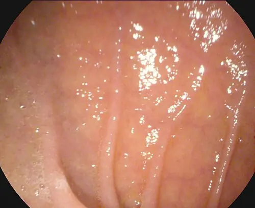
Reduced duodenal folds · endoscopy
- Folds
- Reduction or scalloping
- Surface
- Possible mosaic appearance
- Hypothesis
- Villous atrophy
- Confirmation
- Context, serology, histology
Morphological description
Describe reduced folds, scalloping, fissures and mosaic appearance; involvement may be patchy.
Causes and diagnostic direction
Coeliac disease is an important possibility, but visual markers alone do not determine aetiology.
Differential diagnoses
Other enteropathies cause villous atrophy; serology and histology help distinguish them.
Pitfalls and limitations
Normal endoscopy does not exclude enteropathy; reduced folds do not diagnose coeliac disease.
Take-home message
Describe mucosa, then confirm the suspected cause with appropriate clinical, serological and histological assessment.
Sources
- Celiac Disease: Diagnostic Standards and Dilemmas - PMC ↗
- English | World Gastroenterology Organisation ↗
Documentary review: 2026-10-12
Media source & rights
- Author
- DV Balaban, A Popp, F Vasilescu, D Haidautu, RM Purcarea, M Jinga
- Original source
- DV Balaban, A Popp, F Vasilescu, D Haidautu, RM Purcarea, M Jinga — File : Atrophy of duodenal folds.jpg — CC BY 4.0 — https://commons.wikimedia.org/wiki/File%3AAtrophy_of_duodenal_folds.jpg
- Licence
- CC BY 4.0
- Changes
- EXIF supprimé ; redimensionnement proportionnel ; conversion WebP ; aucun recadrage ni retouche médicale. Dérivé écran proportionnel ≤1280 px, WebP qualité76 ; vignette ≤320 px qualité72. Source acquise et dérivé initial conservés sans modification ; aucune retouche médicale, aucun recadrage.
- Dimensions
- 500 × 407 px
- Check
- 2026-10-11T20:55:37.394532+00:00