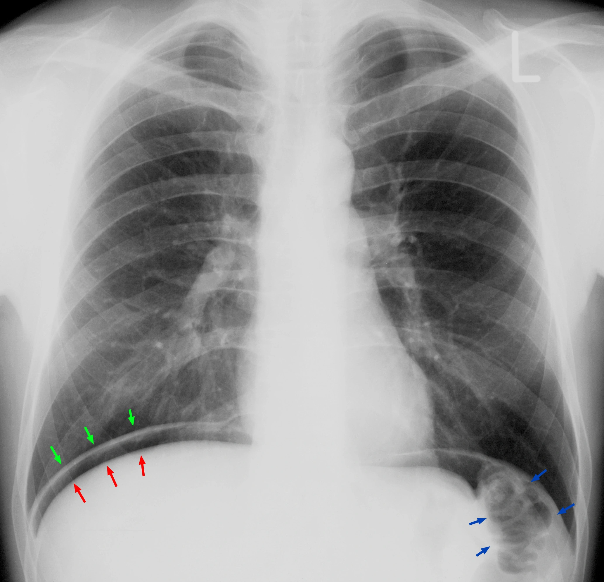
Original MICA image-reading exercise, unscored. No clinical narrative absent from the source is added.
- Describe visible findings before proposing an interpretation.
- Suggest an interpretation and at least one image limitation.
- Which clinical findings or investigations would complement this observation?
Reveal the explained reading
Pneumoperitoneum: subdiaphragmatic free air
- Sign
- Extraluminal gas
- Landmark
- Diaphragm–liver interface
- Further imaging
- CT according to stability
- Priority
- Assess for perforation
Morphological description
The image shows a lucent band beneath the right hemidiaphragm, separate from the bowel gas marked by blue arrows.
Causes and diagnostic direction
Investigate possible bowel perforation. Recent surgery can also account for free gas; the clinical course remains decisive.
Differential diagnoses
Colonic interposition beneath the diaphragm can mimic this sign. Look for haustra and walls; use CT when necessary.
Pitfalls and limitations
A radiograph without visible free air does not exclude perforation. This isolated view cannot localise the bowel source.
Take-home message
Identify free gas, then integrate clinical and procedural context; CT often clarifies the cause, site and complications.
Sources
- Tarasconi et al. Perforated and bleeding peptic ulcer: WSES guidelines (2020) ↗
- Sartelli et al. WSES/GAIS/SIS-E/WSIS/AAST global clinical pathways for patients with intra-abdominal infections (2021) ↗
Documentary review: 2026-10-15
Media source & rights
- Author
- Hellerhoff
- Original source
- Hellerhoff — Pneumoperitoneum.jpg — CC BY-SA 3.0 — https://commons.wikimedia.org/wiki/File:Pneumoperitoneum.jpg
- Licence
- CC BY-SA 3.0
- Changes
- Full original field and annotations retained; WebP conversion, EXIF/GPS metadata removed, proportional derivatives without upscaling, cropping or medical retouching. Derivatives shared under the same CC BY-SA 3.0 licence.
- Dimensions
- 2067 × 1989 px
- Check
- 2026-10-15