Original MICA image-reading exercise, unscored. No clinical narrative absent from the source is added.
- Describe visible findings before proposing an interpretation.
- Suggest an interpretation and at least one image limitation.
- Which clinical findings or investigations would complement this observation?
Reveal the explained reading
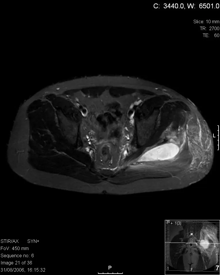
Muscular collection on MRI
- Technique
- MRI
- Observation
- Locate the bright collection and muscular relationships.
- Limitation
- One slice cannot determine the entire extent.
Morphological description
Locate the bright collection and muscular relationships.
Interpretation and diagnostic limits
The source reports sciatic nerve compression.
Differential diagnoses
A collection cannot identify the organism.
Pitfalls
One slice cannot determine the entire extent.
Take-home message
Describe site, contents and anatomical relationships.
Media source & rights
- Author
- Tamer Kamal, Mathew Hall, Ashraf Moharam, Michael Sharr and Jonathan Walczak
- Original source
- Tamer Kamal, Mathew Hall, Ashraf Moharam, Michael Sharr and Jonathan Walczak · IRM : collection musculaire dans une pyomyosite · CC BY 2.0
- Licence
- CC BY 2.0
- Changes
- Full image retained; WebP conversion and proportional resize. No clinical alteration. Derivative retains the source licence.
- Dimensions
- 766 × 959 px
- Check
- 2026-10-14