Original MICA image-reading exercise, unscored. No clinical narrative absent from the source is added.
- Describe visible findings before proposing an interpretation.
- Suggest an interpretation and at least one image limitation.
- Which clinical findings or investigations would complement this observation?
Reveal the explained reading
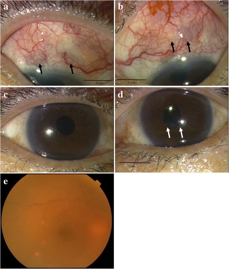
Scleritis and scleral thinning
- Document
- Authentic clinical figure
- Focus
- Scleritis and scleral thinning
- Limitation
- Rheumatoid arthritis does not exclude infection.
Morphological description
Arrows in A–B mark superior scleral abnormalities.
Context and interpretation
The case reports rheumatoid arthritis with ocular inflammation.
Differential diagnoses
Distinguish immune-mediated disease from infection.
Pitfalls and limitations
Rheumatoid arthritis does not exclude infection.
Take-home message
Identify each affected structure.
Sources
- A case of scleritis associated rheumatoid arthritis accompanying an intraocular elevated lesion ↗
- MSD Manual Professional · Red Eye ↗
Documentary review: 2026-10-14
Media source & rights
- Author
- Kobayashi, Takatoshi; Takai, Nanae; Tada, Rei; Shoda, Hiromi; Kida, Teruyo; Ikeda, Tsunehiko; Ozaki, Takurou; Makino, Shigeki
- Original source
- Kobayashi, Takatoshi; Takai, Nanae; Tada, Rei; Shoda, Hiromi; Kida, Teruyo; Ikeda, Tsunehiko; Ozaki, Takurou; Makino, Shigeki · A case of scleritis associated rheumatoid arthritis accompanying an intraocular elevated lesion · CC BY 4.0
- Licence
- CC BY 4.0
- Changes
- Complete original figure retained; WebP conversion and proportional resizing only. No clinical signs added or altered.
- Dimensions
- 788 × 925 px
- Check
- 2026-10-14