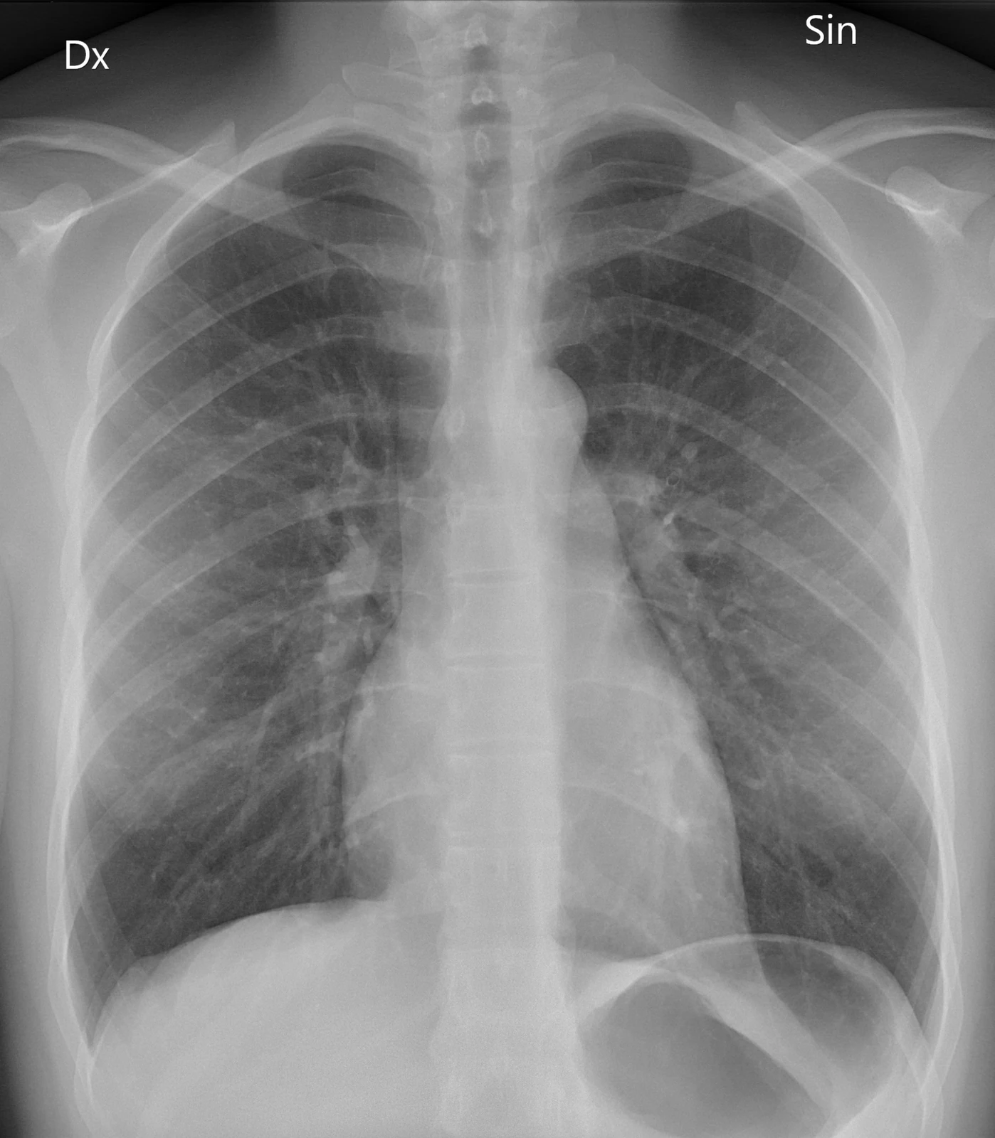
Original MICA image-reading exercise, unscored. No clinical narrative absent from the source is added.
- Describe visible findings before proposing an interpretation.
- Suggest an interpretation and at least one image limitation.
- Which clinical findings or investigations would complement this observation?
Reveal the explained reading
PA chest: reference image
- Technique
- Radiography
- Observation
- Review lungs, hila, mediastinum, heart and diaphragms.
- Limitation
- Radiographic normality does not prove absence of disease.
Morphological description
Review lungs, hila, mediastinum, heart and diaphragms.
Interpretation and diagnostic limits
The source provides a morphological comparison.
Differential diagnoses
A normal radiograph cannot exclude every disease.
Pitfalls
Radiographic normality does not prove absence of disease.
Take-home message
Use a systematic reading method.
Sources
Documentary review: 2026-10-15
Media source & rights
- Author
- Mikael Häggström
- Original source
- Mikael Häggström · Radiographie thoracique de face : comparaison normale · CC0 1.0
- Licence
- CC0 1.0
- Changes
- Full original field and annotations retained; WebP conversion, EXIF/GPS metadata removed, proportional derivatives without upscaling, cropping or medical retouching.
- Dimensions
- 1929 × 2207 px
- Check
- 2026-10-15