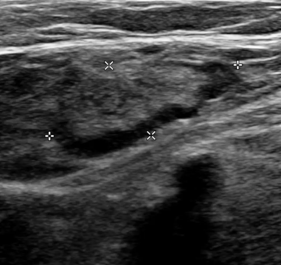
Original MICA image-reading exercise, unscored. No clinical narrative absent from the source is added.
- Describe visible findings before proposing an interpretation.
- Suggest an interpretation and at least one image limitation.
- Which clinical findings or investigations would complement this observation?
Reveal the explained reading
Venous thrombosis · ultrasound
- Major finding
- Non-compressible vein
- B mode
- Lumen and intraluminal material
- Doppler
- Flow and variation
- Interpretation
- Segment and clinical context
Morphological description
Identify the segment, calibre, compressibility and visible material; document the extent examined.
Causes and diagnostic direction
Clinical probability depends on factors including immobility, surgery, cancer and previous thromboembolism.
Differential diagnoses
Artefact and insufficient pressure may mimic abnormality; chronic venous changes can complicate interpretation.
Pitfalls and limitations
A still image cannot demonstrate compression; absent colour flow alone does not confirm thrombosis.
Take-home message
Combine correctly performed compression, venous mapping, Doppler and clinical probability.
Sources
- Point-of-Care Ultrasonography for Deep Venous Thrombosis - StatPearls - NCBI Bookshelf ↗
- Peripheral Venous Duplex Ultrasound: Technique and Clinical Applications - StatPearls - NCBI Bookshelf ↗
Documentary review: 2026-10-12
Media source & rights
- Author
- James Heilman, MD
- Original source
- James Heilman, MD, DVTInGroinNoCompresible.png, CC BY-SA 4.0, Wikimedia Commons; conversion WebP et réduction proportionnelle.
- Licence
- CC BY-SA 4.0
- Changes
- Conversion WebP, redimensionnement proportionnel sans recadrage ni annotations ajoutées; EXIF retiré. Dérivé écran proportionnel ≤1280 px, WebP qualité76 ; vignette ≤320 px qualité72. Source acquise et dérivé initial conservés sans modification ; aucune retouche médicale, aucun recadrage.
- Dimensions
- 960 × 906 px
- Check
- 2026-10-11T20:53:29.467138+00:00