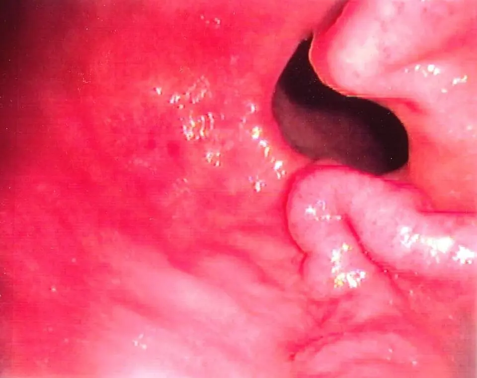
Original MICA image-reading exercise, unscored. No clinical narrative absent from the source is added.
- Describe visible findings before proposing an interpretation.
- Suggest an interpretation and at least one image limitation.
- Which clinical findings or investigations would complement this observation?
Reveal the explained reading
Gastric varices · documented antral location
- Structure
- Dilated gastric veins
- Location
- Describe fundus, body, antrum
- Context
- Portal or segmental hypertension
- Warning
- Gastrointestinal bleeding
Morphological description
Describe submucosal prominence, apparent calibre, extent and surface findings; not every gastric vascular lesion is a varix.
Causes and diagnostic direction
Generalised portal hypertension or splenic venous obstruction can produce gastric varices.
Differential diagnoses
Thickened folds, submucosal tumours and gastric antral vascular ectasia are different diagnoses.
Pitfalls and limitations
A suspected vascular lesion should not be approached as an ordinary mucosal lesion; identify its vascular nature.
Take-home message
Location and vascular anatomy matter as much as appearance; distinguish varices from ectasia.
Sources
Documentary review: 2026-10-12
Media source & rights
- Author
- Samir
- Original source
- Samir — File : Antral varices.jpg — Public domain (release declared by author) — https://commons.wikimedia.org/wiki/File%3AAntral_varices.jpg
- Licence
- Public domain (release declared by author)
- Changes
- EXIF supprimé ; redimensionnement proportionnel ; conversion WebP ; aucun recadrage ni retouche médicale. Dérivé écran proportionnel ≤1280 px, WebP qualité76 ; vignette ≤320 px qualité72. Source acquise et dérivé initial conservés sans modification ; aucune retouche médicale, aucun recadrage.
- Dimensions
- 960 × 761 px
- Check
- 2026-10-11T20:55:37.532937+00:00