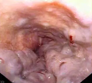
Original MICA image-reading exercise, unscored. No clinical narrative absent from the source is added.
- Describe visible findings before proposing an interpretation.
- Suggest an interpretation and at least one image limitation.
- Which clinical findings or investigations would complement this observation?
Reveal the explained reading
Oesophageal varices · endoscopy
- Structure
- Dilated submucosal veins
- Usual site
- Distal oesophagus
- Description
- Calibre, extent, red markings
- Context
- Portal hypertension
Morphological description
Columns protrude into the lumen; describe red markings without equating them with active bleeding.
Causes and diagnostic direction
Cirrhosis and non-cirrhotic portal hypertension are possible; endoscopic appearance alone does not determine the cause.
Differential diagnoses
Submucosal lesions and folds must be distinguished from vascular columns by complete endoscopic assessment.
Pitfalls and limitations
Varices do not automatically establish cirrhosis; one image cannot describe the complete bleeding risk.
Take-home message
Characterise varices and bleeding signs, then integrate portal and liver disease.
Sources
- Esophageal Varices - StatPearls - NCBI Bookshelf ↗
- Baveno VII – Renewing consensus in portal hypertension - PMC ↗
Documentary review: 2026-10-12
Media source & rights
- Author
- Samir
- Original source
- Samir — File : Esophageal varices - wale.jpg — Public domain (release declared by author) — https://commons.wikimedia.org/wiki/File%3AEsophageal_varices_-_wale.jpg
- Licence
- Public domain (release declared by author)
- Changes
- EXIF supprimé ; redimensionnement proportionnel ; conversion WebP ; aucun recadrage ni retouche médicale. Dérivé écran proportionnel ≤1280 px, WebP qualité76 ; vignette ≤320 px qualité72. Source acquise et dérivé initial conservés sans modification ; aucune retouche médicale, aucun recadrage.
- Dimensions
- 315 × 286 px
- Check
- 2026-10-11T20:55:32.114130+00:00