Flash information
- Cells
- Epithelioid macrophages
- Giant cells
- Possible, not required
- Necrosis
- Assess presence
- Cause
- Infectious or non-infectious
Definition
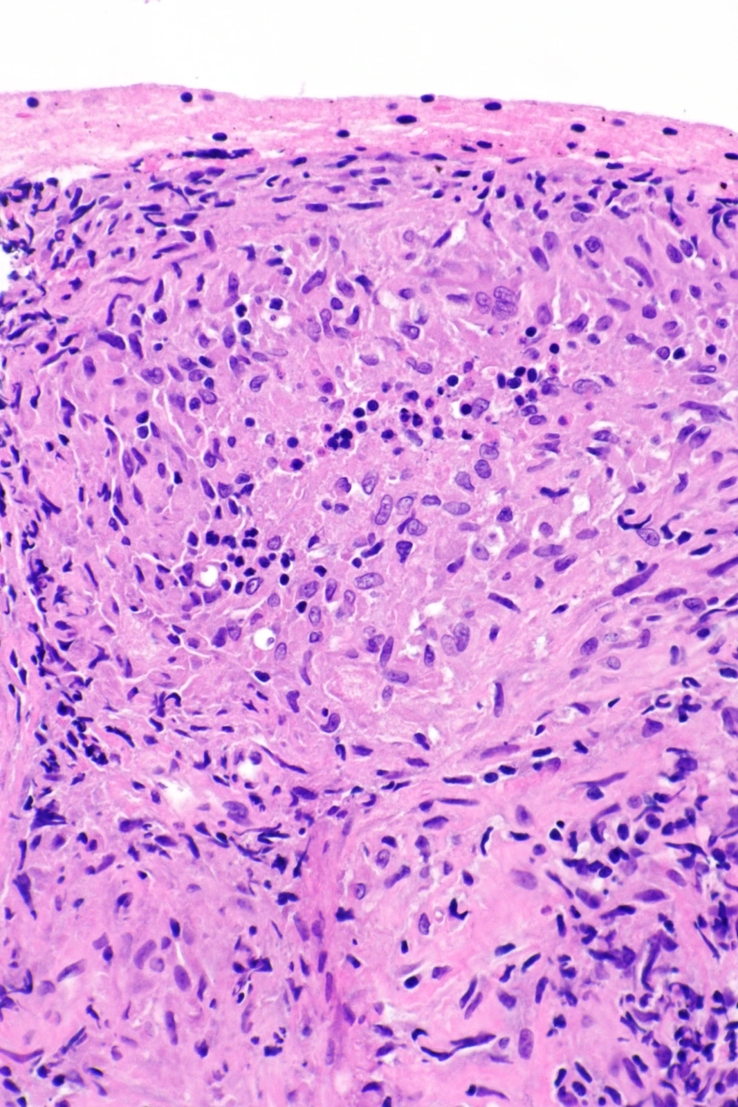
Granulomatous inflammation is a reaction pattern comprising activated macrophage aggregates, often epithelioid.
Examination or reading method
Establish specimen type and stain; in tissue, assess architecture, necrosis and foreign or infectious material.
Morphological description
Describe aggregates, giant cells, peripheral infiltrate and necrosis; aspiration cytology does not show the complete architecture.
Causes and diagnostic direction
Mycobacteria, fungi, foreign material and inflammatory disorders can produce granulomas.
Differential diagnoses
Necrotising and non-necrotising patterns guide but are not specific; infection may also produce non-necrotising granulomas.
Associated findings
Correlate clinical findings, exposure, imaging and microbiological testing; special stains may contribute to investigation.
Red flags
A granuloma does not measure severity; involved organ, possible infection and function determine consequences.
Pitfalls and limitations
A non-necrotising granuloma does not establish sarcoidosis; sampling may miss decisive features.
Take-home message
Identify the pattern, then determine its cause through multidisciplinary correlation.
Sources
- Histopathologic review of granulomatous inflammation - PMC ↗
- Differential diagnosis of granulomatous lung disease: clues and pitfalls: Number 4 in the Series “Pathology for the clinician” Edited by Peter Dorfmüller and Alberto Cavazza - PMC ↗
Documentary review: 2026-10-12
Media source & rights
- Author
- Nephron
- Original source
- Nephron · Granulomes sarcoïdosiques : prélèvement pulmonaire · https://commons.wikimedia.org/wiki/File:Sarcoidosis_-_lung_FNA_--_high_mag.jpg · CC BY-SA 3.0
- Licence
- CC BY-SA 3.0
- Changes
- EXIF omitted; aspect ratio preserved; proportional resize and WebP conversion only. No medical pixels retouched.
- Dimensions
- 1067 × 1600 px
- Check
- 2026-10-11