Flash information
- Document
- Authentic clinical figure
- Focus
- Sectoral episcleritis
- Limitation
- Photographs cannot establish inflammatory depth.
Definition
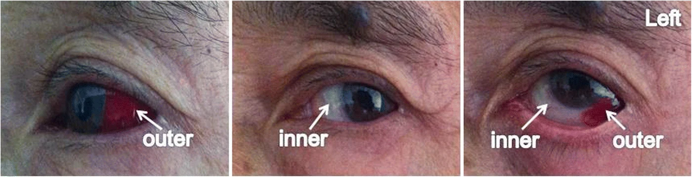
Superficial episcleral inflammation.
Examination or reading method
Assess pain, photophobia and vision.
Morphological description
Sectoral outer-quadrant redness is visible.
Context and interpretation
The source diagnoses episcleritis in this treatment context.
Differential diagnoses
Compare conjunctivitis and scleritis.
Associated findings
Ask about recurrence and extraocular symptoms.
Red flags
Severe pain or visual loss warrants prompt specialist assessment.
Pitfalls and limitations
Photographs cannot establish inflammatory depth.
Take-home message
Describe redness before assigning a cause.
Sources
- Episcleritis in a patient with mucosal melanoma treated with interferon alfa-2b and radiotherapy: a case report ↗
- MSD Manual Professional · Red Eye ↗
Documentary review: 2026-10-14
Media source & rights
- Author
- Yang, Li; Ji, Shaofei; Wang, Liping; Zhang, Yi
- Original source
- Yang, Li; Ji, Shaofei; Wang, Liping; Zhang, Yi · Episcleritis in a patient with mucosal melanoma treated with interferon alfa-2b and radiotherapy: a case report · CC BY 4.0
- Licence
- CC BY 4.0
- Changes
- Complete original figure retained; WebP conversion and proportional resizing only. No clinical signs added or altered.
- Dimensions
- 1400 × 359 px
- Check
- 2026-10-14