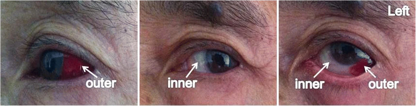
Original MICA image-reading exercise, unscored. No clinical narrative absent from the source is added.
- Describe visible findings before proposing an interpretation.
- Suggest an interpretation and at least one image limitation.
- Which clinical findings or investigations would complement this observation?
Reveal the explained reading
Sectoral episcleritis
- Document
- Authentic clinical figure
- Focus
- Sectoral episcleritis
- Limitation
- Photographs cannot establish inflammatory depth.
Morphological description
Sectoral outer-quadrant redness is visible.
Context and interpretation
The source diagnoses episcleritis in this treatment context.
Differential diagnoses
Compare conjunctivitis and scleritis.
Pitfalls and limitations
Photographs cannot establish inflammatory depth.
Take-home message
Describe redness before assigning a cause.
Sources
- Episcleritis in a patient with mucosal melanoma treated with interferon alfa-2b and radiotherapy: a case report ↗
- MSD Manual Professional · Red Eye ↗
Documentary review: 2026-10-14
Media source & rights
- Author
- Yang, Li; Ji, Shaofei; Wang, Liping; Zhang, Yi
- Original source
- Yang, Li; Ji, Shaofei; Wang, Liping; Zhang, Yi · Episcleritis in a patient with mucosal melanoma treated with interferon alfa-2b and radiotherapy: a case report · CC BY 4.0
- Licence
- CC BY 4.0
- Changes
- Complete original figure retained; WebP conversion and proportional resizing only. No clinical signs added or altered.
- Dimensions
- 1400 × 359 px
- Check
- 2026-10-14