Flash information
- Technique
- Doppler ultrasound
- Observation
- Assess wall thickening around the lumen.
- Limitation
- Doppler settings alter signal appearance.
Definition
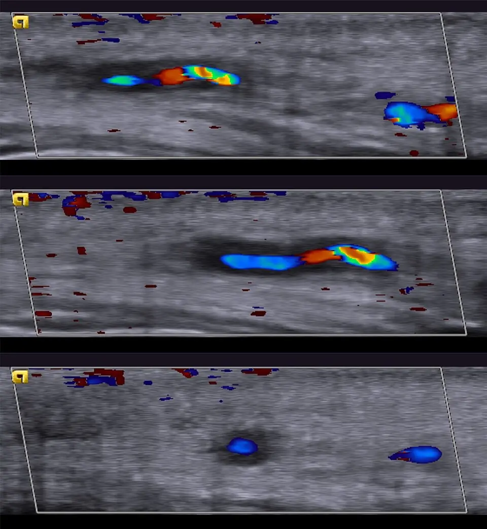
The author identifies temporal arteritis on ultrasound.
Reading method
Compare lumen, wall and colour signal across three views.
Morphological description
Assess wall thickening around the lumen.
Interpretation and diagnostic limits
Diagnosis requires clinical context.
Differential diagnoses
Distinguish inflammatory thickening from other wall abnormalities.
Clinical correlation
Assess new headache, jaw claudication and visual symptoms.
Clinical impact
Visual symptoms with suspected arteritis are urgent.
Pitfalls
Doppler settings alter signal appearance.
Take-home message
Colour alone does not establish diagnosis.
Sources
Documentary review: 2026-10-14
Media source & rights
- Author
- Hellerhoff
- Original source
- Hellerhoff · Doppler : artérite temporale décrite dans la source · CC BY-SA 4.0
- Licence
- CC BY-SA 4.0
- Changes
- Full image retained; WebP conversion and proportional resize. No clinical alteration. Derivative retains the source licence.
- Dimensions
- 960 × 1042 px
- Check
- 2026-10-14