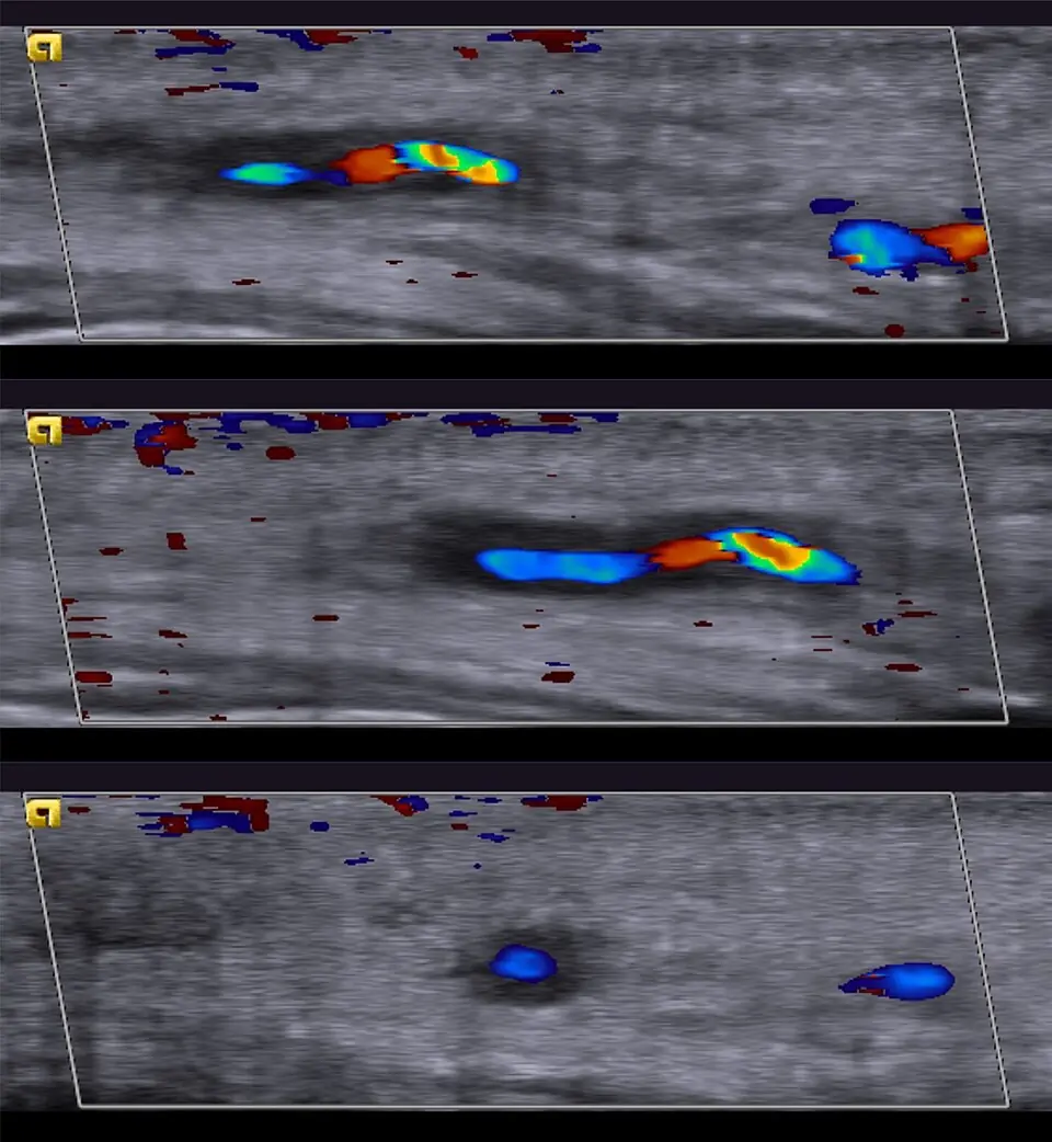
Original MICA image-reading exercise, unscored. No clinical narrative absent from the source is added.
- Describe visible findings before proposing an interpretation.
- Suggest an interpretation and at least one image limitation.
- Which clinical findings or investigations would complement this observation?
Reveal the explained reading
Temporal artery wall on Doppler
- Technique
- Doppler ultrasound
- Observation
- Assess wall thickening around the lumen.
- Limitation
- Doppler settings alter signal appearance.
Morphological description
Assess wall thickening around the lumen.
Interpretation and diagnostic limits
Diagnosis requires clinical context.
Differential diagnoses
Distinguish inflammatory thickening from other wall abnormalities.
Pitfalls
Doppler settings alter signal appearance.
Take-home message
Colour alone does not establish diagnosis.
Sources
Documentary review: 2026-10-14
Media source & rights
- Author
- Hellerhoff
- Original source
- Hellerhoff · Doppler : artérite temporale décrite dans la source · CC BY-SA 4.0
- Licence
- CC BY-SA 4.0
- Changes
- Full image retained; WebP conversion and proportional resize. No clinical alteration. Derivative retains the source licence.
- Dimensions
- 960 × 1042 px
- Check
- 2026-10-14