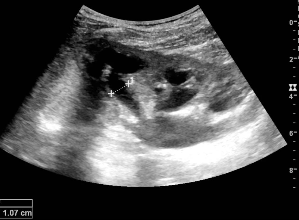
Flash information
- Structure
- Pelvis and calyces
- Appearance
- Communicating anechoic spaces
- Parenchyma
- Assess thickness
- Cause
- Assess obstruction in context
Definition
Hydronephrosis is renal collecting-system dilatation; ultrasound alone does not identify the obstruction.
Examination or reading method
Scan both kidneys in multiple planes, renal parenchyma and bladder; assess visible causes.
Morphological description
Describe pelvic and calyceal dilatation, laterality and possible cortical thinning.
Causes and diagnostic direction
Stones, tumours, narrowing or bladder dysfunction can cause dilatation; some situations are non-obstructive.
Differential diagnoses
Non-communicating parapelvic cysts and an extrarenal pelvis may mimic dilatation.
Associated findings
Relate findings to pain, urine output, renal function and infection; investigate further if cause remains unclear.
Red flags
Fever with suspected obstruction, anuria or renal impairment requires urgent assessment.
Pitfalls and limitations
Dilatation is not absolute proof of obstruction; assess continuity of spaces and clinical context.
Take-home message
Describe the collecting system and parenchyma, then determine cause and clinical consequences.
Sources
- Hydronephrosis and Hydroureter - StatPearls - NCBI Bookshelf ↗
- Point of care renal ultrasonography for the busy nephrologist: A pictorial review - PMC ↗
Documentary review: 2026-10-12
Media source & rights
- Author
- Kristoffer Lindskov Hansen; Michael Bachmann Nielsen; Caroline Ewertsen
- Original source
- Kristoffer Lindskov Hansen; Michael Bachmann Nielsen; Caroline Ewertsen · Échographie : dilatation pyélocalicielle documentée · https://commons.wikimedia.org/wiki/File:Ultrasonography_of_hydronephrosis_with_dilated_anechoic_pelvis_and_calyces,_along_with_cortical_atrophy.jpg · CC BY 4.0
- Licence
- CC BY 4.0
- Changes
- EXIF omitted; aspect ratio preserved; proportional resize and WebP conversion only. No medical pixels retouched.
- Dimensions
- 1006 × 741 px
- Check
- 2026-10-11