 Images
MICA IMAGES A0057
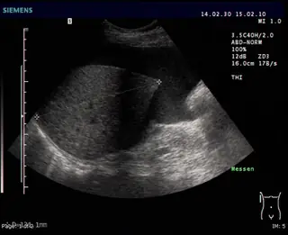
Ascites on ultrasound
Free intraperitoneal fluid.
Drahreg01 · Ascite à l’échographie · CC BY-SA 3.0
Open accessOpen
MICA
Communicating fluid spaces in the pelvis and calyces; their cause and obstructive significance require assessment.
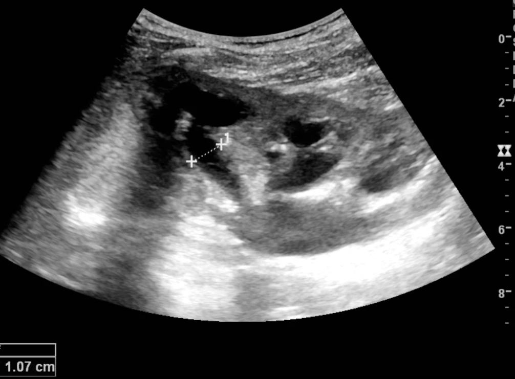
Kristoffer Lindskov Hansen; Michael Bachmann Nielsen; Caroline Ewertsen · Échographie : dilatation pyélocalicielle documentée · https://commons.wikimedia.org/wiki/File:Ultrasonography_of_hydronephrosis_with_dilated_anechoic_pelvis_and_calyces,_along_with_cortical_atrophy.jpg · CC BY 4.0
Kristoffer Lindskov Hansen; Michael Bachmann Nielsen; Caroline Ewertsen
View the original sourceTeaching notes based on the cited sources.
ImagesMICA IMAGES A0057
Free intraperitoneal fluid.
Drahreg01 · Ascite à l’échographie · CC BY-SA 3.0
 Images
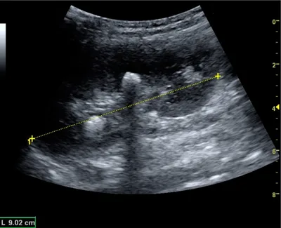
MICA IMAGES A0087
Central stone with posterior acoustic shadowing.
Kristoffer Lindskov Hansen, Michael Bachmann Nielsen and Caroline Ewertsen · Calcul rénal avec ombre acoustique · CC BY 4.0
 Images
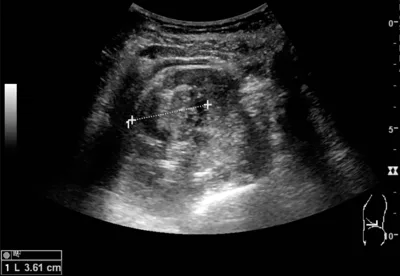
MICA IMAGES A0088
A solid cortical renal mass on ultrasound.
Kristoffer Lindskov Hansen, Michael Bachmann Nielsen and Caroline Ewertsen · Masse rénale solide : échographie · CC BY 4.0
Learning objective
Observe and describe the document, then compare your interpretation with the atlas sheet.
Points to observe
- Scan both kidneys in multiple planes, renal parenchyma and bladder; assess visible causes.
Open observation mode