Flash information
- Document
- Authentic MRI
- Focus
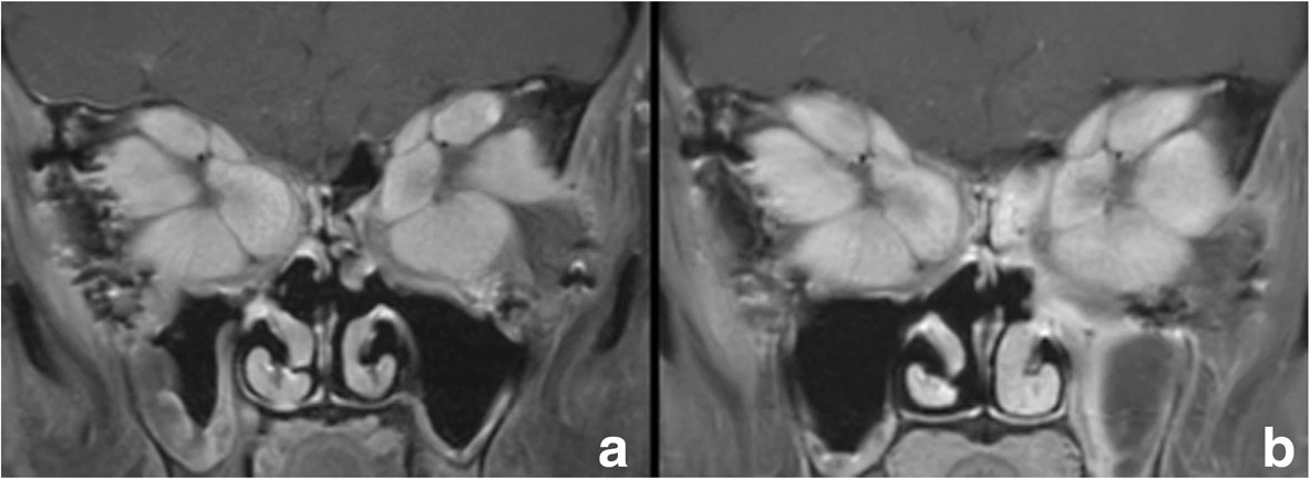
- MRI: extraocular muscles in Graves orbitopathy
- Limitation
- This figure cannot individually quantify disease activity.
Definition
Orbital muscular involvement in a severe Graves case.
Examination or reading method
Compare muscles and orbital apices between panels.
Morphological description
The source documents enlarged extraocular muscles.
Context and interpretation
The case reports compression affecting optic nerves.
Differential diagnoses
Consider other orbital inflammation and masses.
Associated findings
Correlate visual function and motility.
Red flags
Suspected optic neuropathy warrants urgent care.
Pitfalls and limitations
This figure cannot individually quantify disease activity.
Take-home message
Combine imaging and clinical examination.
Sources
Documentary review: 2026-10-15
Media source & rights
- Author
- Rau, Andrea; Klopfer, Matthias; Rommel, Niklas; Rau-Fornefeld, Mechthild; Kolk, Andreas
- Original source
- Rau, Andrea; Klopfer, Matthias; Rommel, Niklas; Rau-Fornefeld, Mechthild; Kolk, Andreas · Extraocular muscle repositioning as the last therapeutic option for a patient with a severe course of Graves’ Ophthalmopathy: a case report · CC BY 4.0 · https://link.springer.com/article/10.1186/s12886-018-0718-1
- Licence
- CC BY 4.0
- Changes
- Full original field and annotations retained; WebP conversion, EXIF/GPS metadata removed, proportional derivatives without upscaling, cropping or medical retouching.
- Dimensions
- 1183 × 433 px
- Check
- 2026-10-15