Original MICA image-reading exercise, unscored. No clinical narrative absent from the source is added.
- Describe visible findings before proposing an interpretation.
- Suggest an interpretation and at least one image limitation.
- Which clinical findings or investigations would complement this observation?
Reveal the explained reading
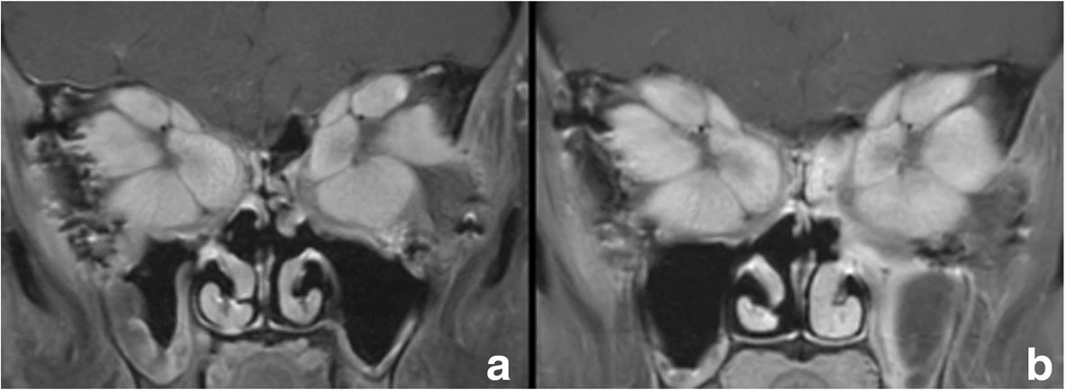
MRI: extraocular muscles in Graves orbitopathy
- Document
- Authentic MRI
- Focus
- MRI: extraocular muscles in Graves orbitopathy
- Limitation
- This figure cannot individually quantify disease activity.
Morphological description
The source documents enlarged extraocular muscles.
Context and interpretation
The case reports compression affecting optic nerves.
Differential diagnoses
Consider other orbital inflammation and masses.
Pitfalls and limitations
This figure cannot individually quantify disease activity.
Take-home message
Combine imaging and clinical examination.
Sources
Documentary review: 2026-10-15
Media source & rights
- Author
- Rau, Andrea; Klopfer, Matthias; Rommel, Niklas; Rau-Fornefeld, Mechthild; Kolk, Andreas
- Original source
- Rau, Andrea; Klopfer, Matthias; Rommel, Niklas; Rau-Fornefeld, Mechthild; Kolk, Andreas · Extraocular muscle repositioning as the last therapeutic option for a patient with a severe course of Graves’ Ophthalmopathy: a case report · CC BY 4.0 · https://link.springer.com/article/10.1186/s12886-018-0718-1
- Licence
- CC BY 4.0
- Changes
- Full original field and annotations retained; WebP conversion, EXIF/GPS metadata removed, proportional derivatives without upscaling, cropping or medical retouching.
- Dimensions
- 1183 × 433 px
- Check
- 2026-10-15