PET
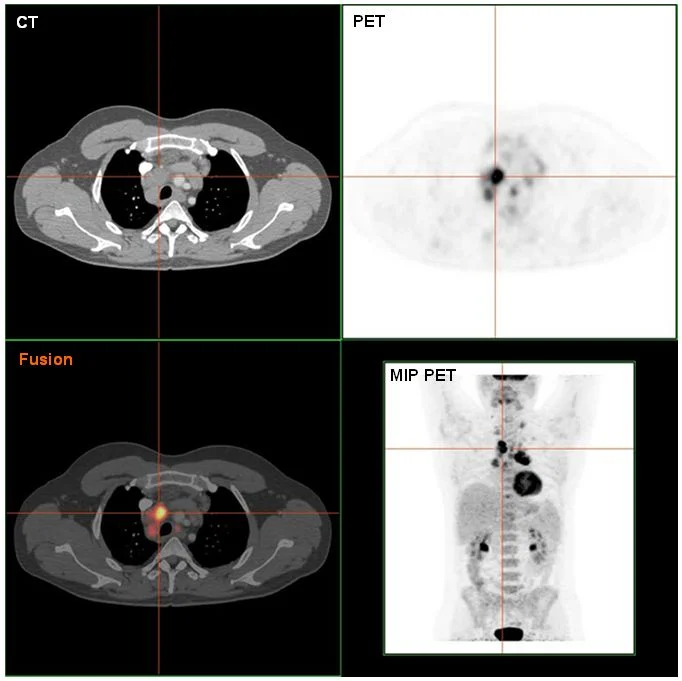
Lymphoma: fused PET-CT
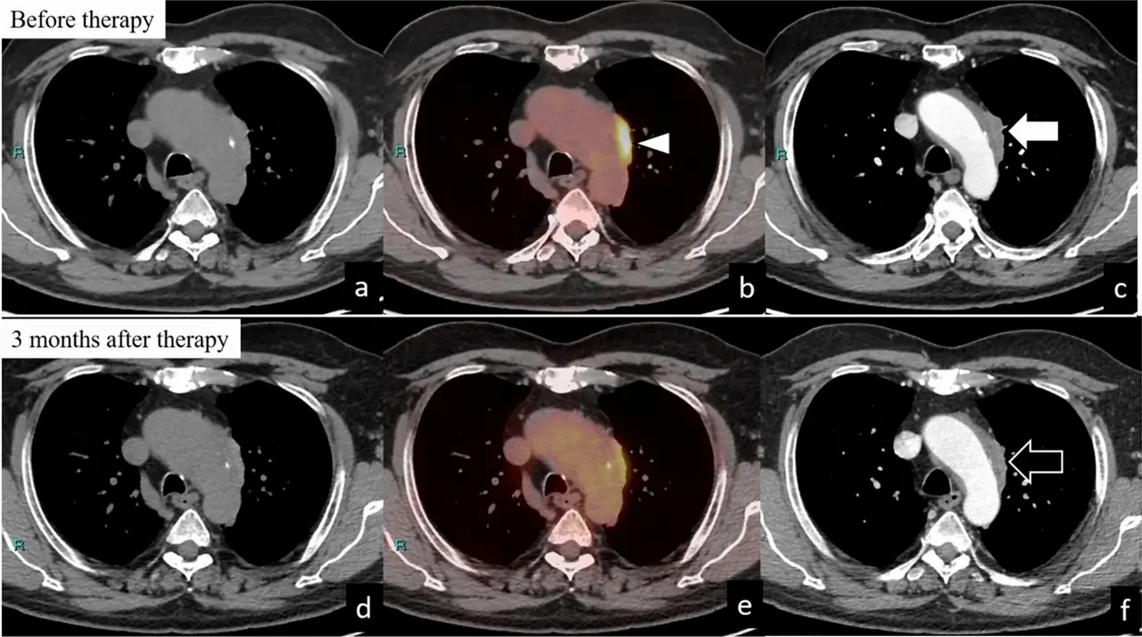
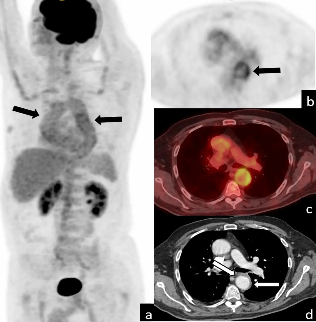
PET-CT of source-confirmed Hodgkin lymphoma.
- Observation
- An intensely avid region appears in the image.
- Limitation
- The palette cannot replace quantitative data.
MICA
Observe the images and explore this collection’s sheets.
← Morphological semiology & imaging3 sheets