Flash information
- Observation
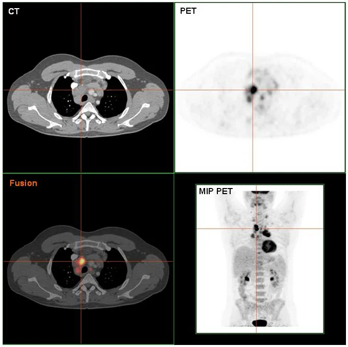
- An intensely avid region appears in the image.
- Limitation
- The palette cannot replace quantitative data.
Definition
PET-CT of source-confirmed Hodgkin lymphoma.
Reading method
Compare anatomical and metabolic information.
Morphological description
An intensely avid region appears in the image.
Source context
Histological confirmation belongs to the source case.
Differential diagnoses
Inflammation can also show FDG uptake.
Clinical correlation
Correlate biopsy and complete staging.
Clinical impact
One slice cannot establish stage.
Pitfalls and limitations
The palette cannot replace quantitative data.
Take-home message
Uptake is not histological proof.
Media source & rights
- Author
- Hg6996
- Original source
- Hg6996 · Lymphome : TEP-TDM fusionnée · Public domain
- Licence
- Public domain
- Changes
- Whole image retained; proportional resize and WebP conversion only. No clinical alteration. Derivative retains the original licence.
- Dimensions
- 682 × 681 px
- Check
- 2026-10-14