Original MICA image-reading exercise, unscored. No clinical narrative absent from the source is added.
- Describe visible findings before proposing an interpretation.
- Suggest an interpretation and at least one image limitation.
- Which clinical findings or investigations would complement this observation?
Reveal the explained reading
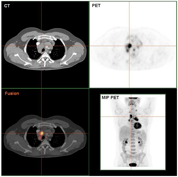
Lymphoma: fused PET-CT
- Observation
- An intensely avid region appears in the image.
- Limitation
- The palette cannot replace quantitative data.
Morphological description
An intensely avid region appears in the image.
Source context
Histological confirmation belongs to the source case.
Differential diagnoses
Inflammation can also show FDG uptake.
Pitfalls and limitations
The palette cannot replace quantitative data.
Take-home message
Uptake is not histological proof.
Media source & rights
- Author
- Hg6996
- Original source
- Hg6996 · Lymphome : TEP-TDM fusionnée · Public domain
- Licence
- Public domain
- Changes
- Whole image retained; proportional resize and WebP conversion only. No clinical alteration. Derivative retains the original licence.
- Dimensions
- 682 × 681 px
- Check
- 2026-10-14