View the original document
ImagesTEACHING RESOURCE M0003
HEp-2 fluorescence patterns: ICAP
An ICAP consensus figure showing several nuclear and cytoplasmic fluorescence patterns on HEp-2 cells.
ICAP · Frontiers
Free MICA accountOpen
MICA
Cutaneous small-vessel involvement with neutrophils, nuclear debris and wall injury.
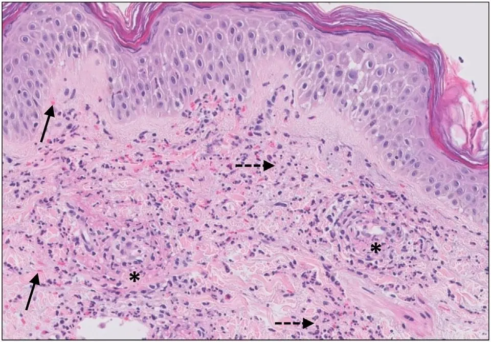
Jenny Giang, Marc A. J. Seelen, Martijn B. A. van Doorn, Robert Rissmann,Errol P. Prens and Jeffrey Damman — File : Micrograph of cutaneous small-vessel vasculitis.jpg — CC BY 4.0 — https://commons.wikimedia.org/wiki/File%3AMicrograph_of_cutaneous_small-vessel_vasculitis.jpg
Jenny Giang, Marc A. J. Seelen, Martijn B. A. van Doorn, Robert Rissmann,Errol P. Prens and Jeffrey Damman
View the original sourceTeaching notes based on the cited sources.
TEACHING RESOURCE M0003
An ICAP consensus figure showing several nuclear and cytoplasmic fluorescence patterns on HEp-2 cells.
ICAP · Frontiers
TEACHING RESOURCE M0004
An interview with Professor Zahir Amoura on when to request antinuclear antibodies and how to interpret them.
Fréquence Médicale
 Images
MICA IMAGES A0038
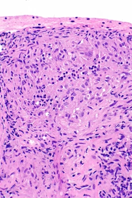
A group of epithelioid macrophages, sometimes with giant cells; this pattern does not define one cause.
Nephron · Granulomes sarcoïdosiques : prélèvement pulmonaire · https://commons.wikimedia.org/wiki/File:Sarcoidosis_-_lung_FNA_--_high_mag.jpg · CC BY-SA 3.0
Learning objective
Observe and describe the document, then compare your interpretation with the atlas sheet.
Points to observe
- Establish biopsy lesion age and site; examine vessel walls and consider immunofluorescence when indicated.
Open observation mode