View the original document
ImagesTEACHING RESOURCE M0003
HEp-2 fluorescence patterns: ICAP
An ICAP consensus figure showing several nuclear and cytoplasmic fluorescence patterns on HEp-2 cells.
ICAP · Frontiers
Free MICA accountOpen
MICA
Small fibres at fascicle edges: distribution is essential.
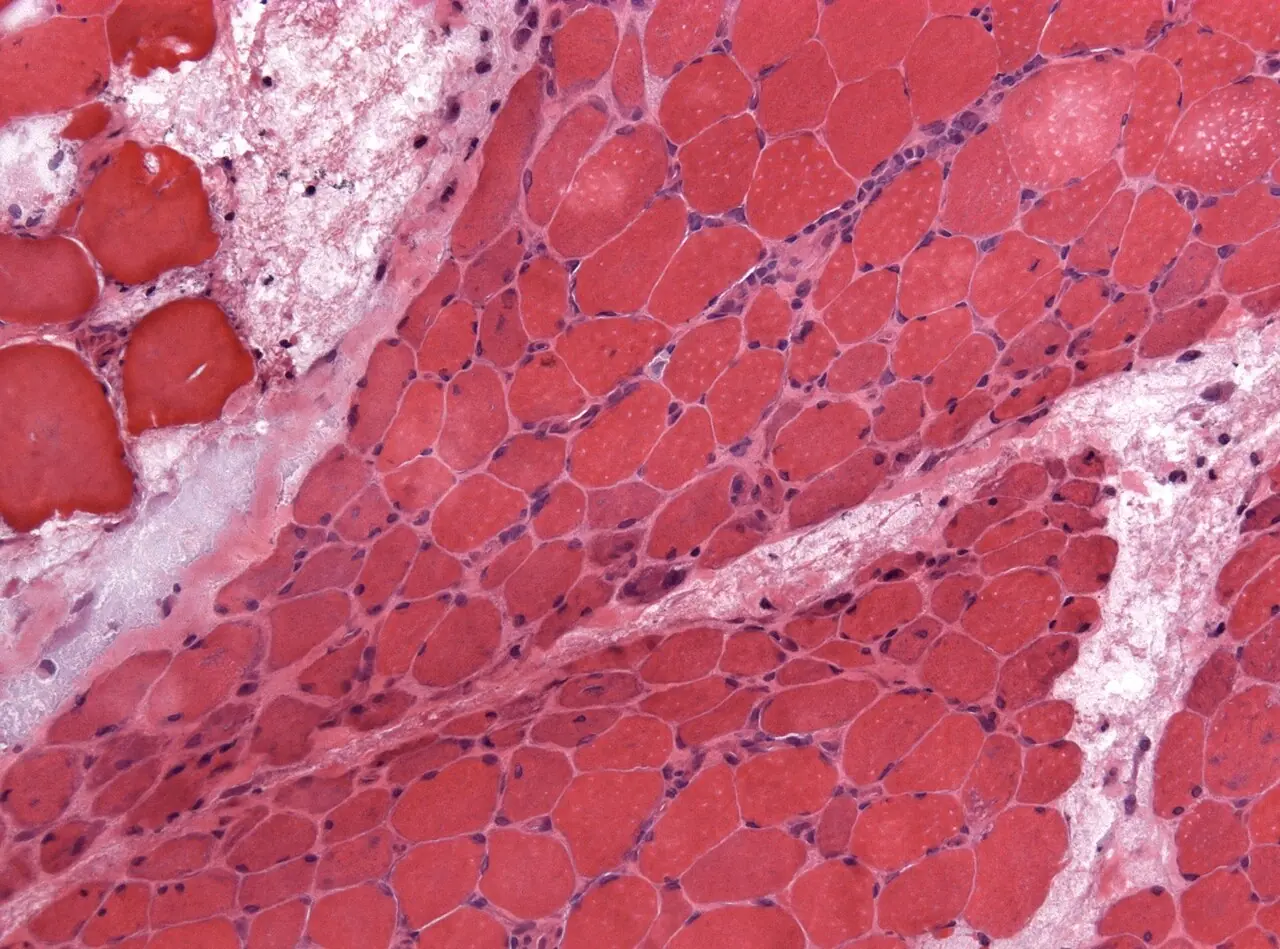
Jensflorian — File : NP MGMT 0252.jpg — CC BY-SA 4.0 — https://commons.wikimedia.org/wiki/File%3ANP_MGMT_0252.jpg
Jensflorian
View the original sourceTeaching notes based on the cited sources.
TEACHING RESOURCE M0003
An ICAP consensus figure showing several nuclear and cytoplasmic fluorescence patterns on HEp-2 cells.
ICAP · Frontiers
TEACHING RESOURCE M0004
An interview with Professor Zahir Amoura on when to request antinuclear antibodies and how to interpret them.
Fréquence Médicale
 Images
MICA IMAGES A0038
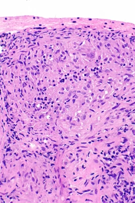
A group of epithelioid macrophages, sometimes with giant cells; this pattern does not define one cause.
Nephron · Granulomes sarcoïdosiques : prélèvement pulmonaire · https://commons.wikimedia.org/wiki/File:Sarcoidosis_-_lung_FNA_--_high_mag.jpg · CC BY-SA 3.0
Learning objective
Observe and describe the document, then compare your interpretation with the atlas sheet.
Points to observe
- Compare fascicle centres and edges; check specimen orientation and quality.
Open observation mode