View the original document
ImagesTEACHING RESOURCE M0003
HEp-2 fluorescence patterns: ICAP
An ICAP consensus figure showing several nuclear and cytoplasmic fluorescence patterns on HEp-2 cells.
ICAP · Frontiers
Free MICA accountOpen
MICA
Four steps connect phenotype, sampling, differential diagnoses and synthesis. No microscopic tissue is artificially reconstructed.
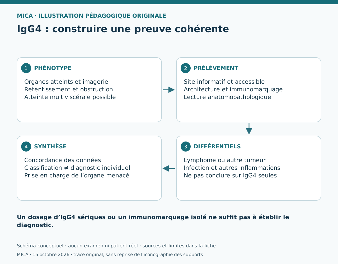
MICA · schéma original du 15 octobre 2026 ; aucune image tierce incorporée
MICA · composition vectorielle originale
View the original sourceCréation originale pour MICA · diffusion dans le campus autorisée par la demande du 15 octobre 2026
Teaching notes based on the cited sources.
TEACHING RESOURCE M0003
An ICAP consensus figure showing several nuclear and cytoplasmic fluorescence patterns on HEp-2 cells.
ICAP · Frontiers
TEACHING RESOURCE M0004
An interview with Professor Zahir Amoura on when to request antinuclear antibodies and how to interpret them.
Fréquence Médicale
 Images
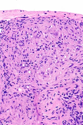
MICA IMAGES A0038
A group of epithelioid macrophages, sometimes with giant cells; this pattern does not define one cause.
Nephron · Granulomes sarcoïdosiques : prélèvement pulmonaire · https://commons.wikimedia.org/wiki/File:Sarcoidosis_-_lung_FNA_--_high_mag.jpg · CC BY-SA 3.0
Learning objective
Observe and describe the document, then compare your interpretation with the atlas sheet.
Points to observe
- Read the cards and compare them with the clinical context and sources. Numbers are reading guides without quantitative weights.
Open observation mode