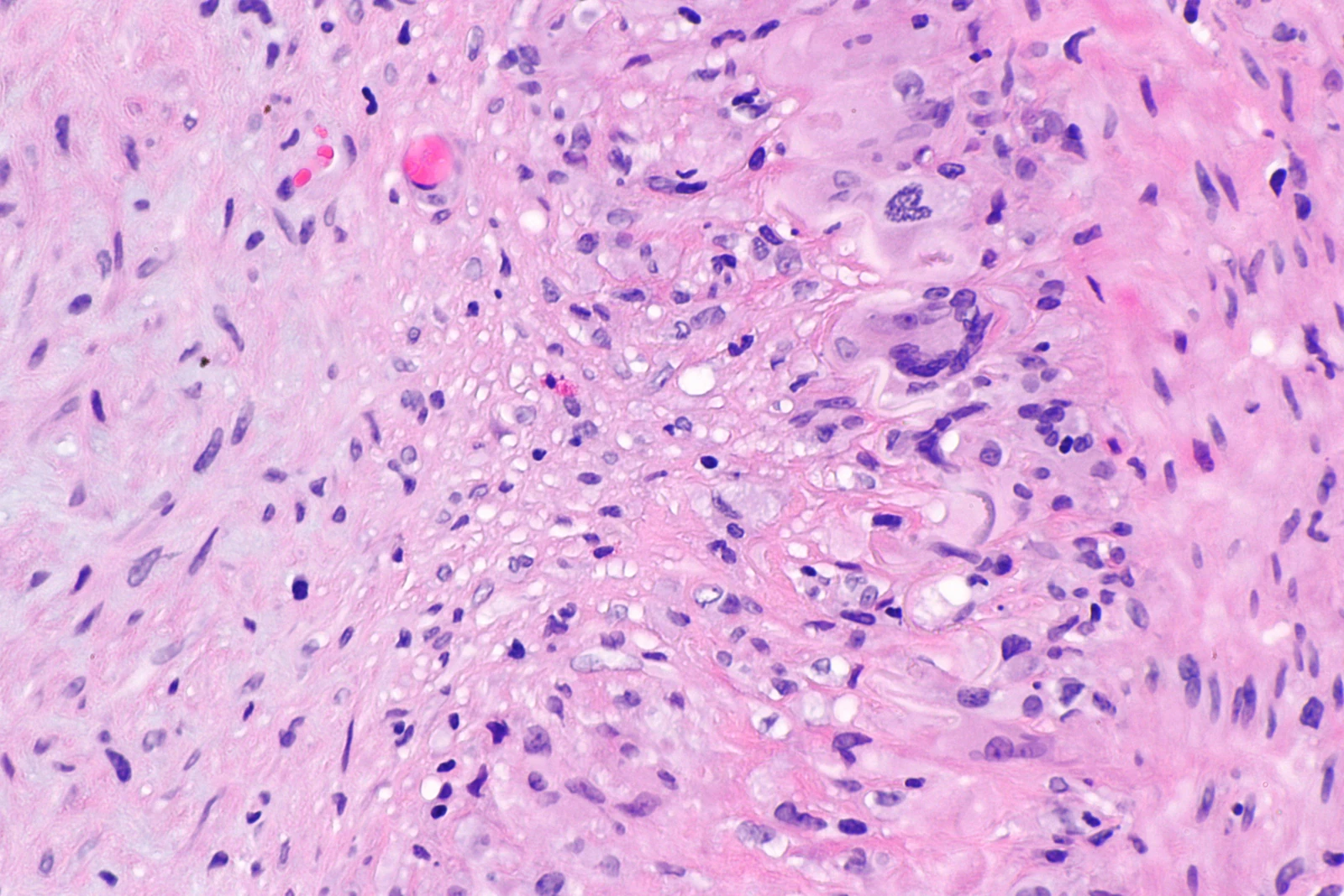
Original MICA image-reading exercise, unscored. No clinical narrative absent from the source is added.
- Describe visible findings before proposing an interpretation.
- Suggest an interpretation and at least one image limitation.
- Which clinical findings or investigations would complement this observation?
Reveal the explained reading
Giant cell arteritis · histology
- Vessel
- Artery
- Inflammation
- Arterial wall
- Elastic layer
- Assess disruption
- Sampling
- Possible skip lesions
Morphological description
Assess wall infiltrate, elastic-layer damage, intimal hyperplasia and possible giant cells.
Causes and diagnostic direction
This pattern may support diagnosis in a compatible presentation; giant cells are not required.
Differential diagnoses
Distinguish normal artery, limited inflammation and other vascular lesions; architecture and supplementary stains help.
Pitfalls and limitations
Lesions may be segmental; a negative biopsy does not exclude arteritis, particularly with limited sampling.
Take-home message
Interpret the specimen with clinical findings and imaging, accounting for sampling limitations.
Sources
- Giant Cell Arteritis (Temporal Arteritis) - StatPearls - NCBI Bookshelf ↗
- Temporal Artery Biopsy - StatPearls - NCBI Bookshelf ↗
Documentary review: 2026-10-15
Media source & rights
- Author
- Nephron
- Original source
- Nephron — File : Giant cell arteritis -- high mag.jpg — CC BY-SA 3.0 — https://commons.wikimedia.org/wiki/File%3AGiant_cell_arteritis_--_high_mag.jpg
- Licence
- CC BY-SA 3.0
- Changes
- Full original field and annotations retained; WebP conversion, EXIF/GPS metadata removed, proportional derivatives without upscaling, cropping or medical retouching. Derivatives shared under the same CC BY-SA 3.0 licence.
- Dimensions
- 4272 × 2848 px
- Check
- 2026-10-15