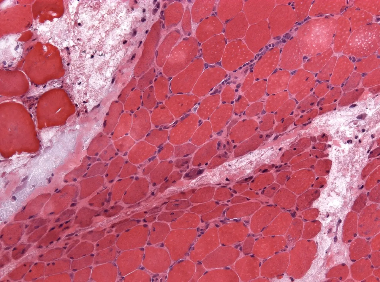
Original MICA image-reading exercise, unscored. No clinical narrative absent from the source is added.
- Describe visible findings before proposing an interpretation.
- Suggest an interpretation and at least one image limitation.
- Which clinical findings or investigations would complement this observation?
Reveal the explained reading
Perifascicular atrophy · muscle biopsy
- Tissue
- Skeletal muscle
- Distribution
- Fascicle periphery
- Morphology
- Reduced fibre diameter
- Direction
- Dermatomyositis, in context
Morphological description
Describe peripheral small fibres, inflammation, necrosis and associated changes.
Causes and diagnostic direction
This pattern supports dermatomyositis, but not every dermatomyositis biopsy shows it.
Differential diagnoses
Diffuse or grouped atrophy has a different distribution; complete characterisation requires several analyses.
Pitfalls and limitations
An unrepresentative biopsy may miss the pattern; morphology must be correlated clinically.
Take-home message
Recognise the perifascicular distribution and integrate other pathological, immunological and clinical findings.
Sources
- Muscle biopsy features of idiopathic inflammatory myopathies and differential diagnosis - PMC ↗
- Dermatomyositis: Muscle Pathology According to Antibody Subtypes - PubMed ↗
Documentary review: 2026-10-12
Media source & rights
- Author
- Jensflorian
- Original source
- Jensflorian — File : NP MGMT 0252.jpg — CC BY-SA 4.0 — https://commons.wikimedia.org/wiki/File%3ANP_MGMT_0252.jpg
- Licence
- CC BY-SA 4.0
- Changes
- EXIF supprimé ; redimensionnement proportionnel ; conversion WebP ; aucun recadrage ni retouche médicale. Dérivé écran proportionnel ≤1280 px, WebP qualité76 ; vignette ≤320 px qualité72. Source acquise et dérivé initial conservés sans modification ; aucune retouche médicale, aucun recadrage.
- Dimensions
- 1280 × 949 px
- Check
- 2026-10-11T20:56:53.282695+00:00