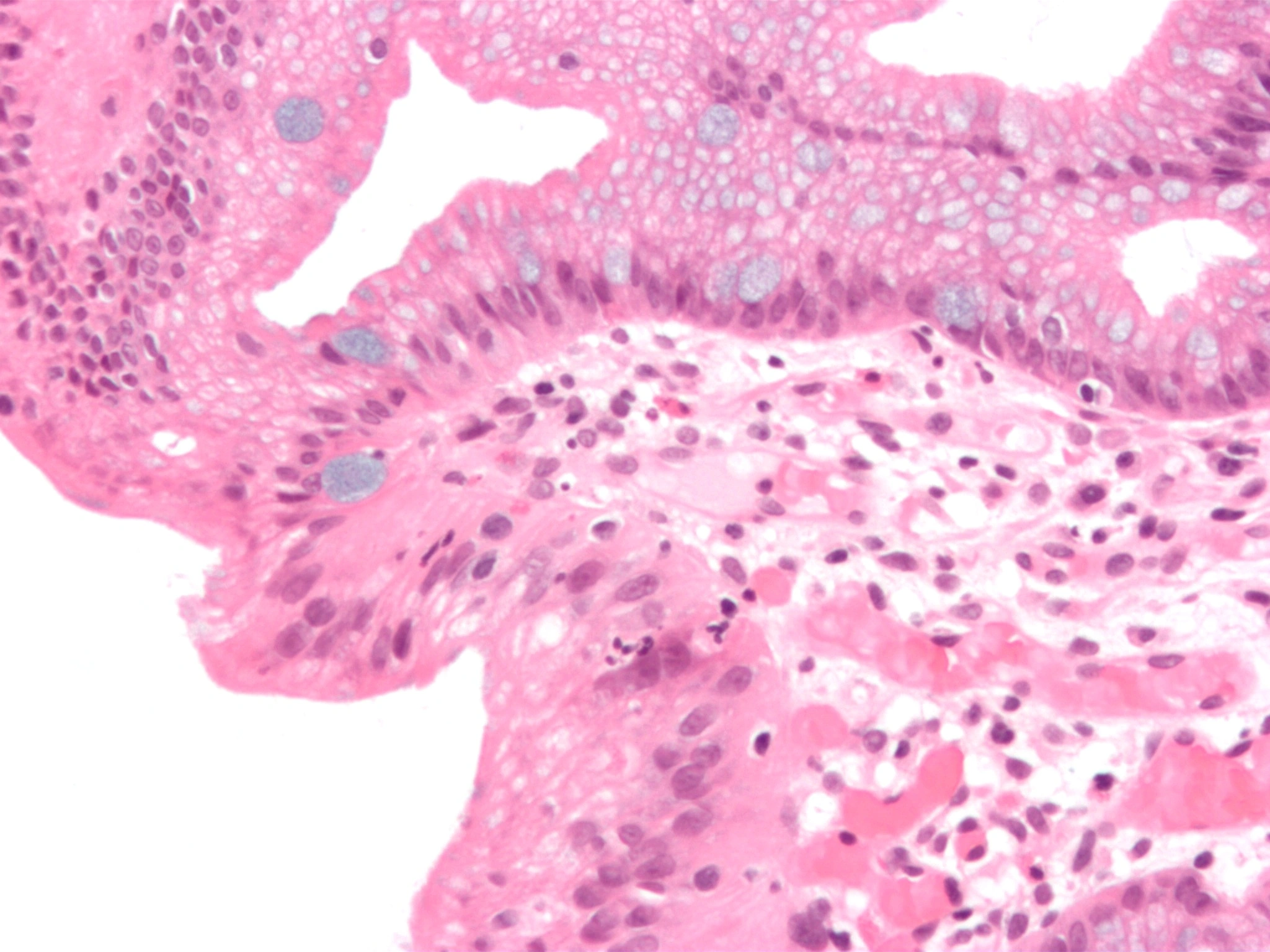
Original MICA image-reading exercise, unscored. No clinical narrative absent from the source is added.
- Describe visible findings before proposing an interpretation.
- Suggest an interpretation and at least one image limitation.
- Which clinical findings or investigations would complement this observation?
Reveal the explained reading
Oesophageal intestinal metaplasia · Alcian blue
- Technique
- Histology
- Stain
- Alcian blue
- Clue
- Goblet cells
- Limit
- Metaplasia ≠ cancer
Morphological description
Goblet cells contain blue-stained mucins; the field shows glandular epithelium.
Causes and diagnostic direction
Chronic gastro-oesophageal reflux is a common setting; the section alone cannot establish the history.
Differential diagnoses
Cardia intestinal metaplasia requires localisation for distinction; blue staining does not establish dysplasia.
Pitfalls and limitations
Metaplasia, dysplasia and adenocarcinoma are distinct; dysplasia requires expert pathological confirmation.
Take-home message
Histology characterises tissue; surveillance depends on the complete diagnosis rather than blue cells alone.
Sources
- Weusten BLAM et al. Diagnosis and management of Barrett esophagus: ESGE Guideline. Endoscopy, 2023 ↗
- Shaheen NJ. Diagnosis and Management of Barrett’s Esophagus: An Updated ACG Guideline. ACG Virtual Grand Rounds, 28 July 2022(présentation de l’auteur, pas journal article) ↗
Documentary review: 2026-10-15
Media source & rights
- Author
- Nephron
- Original source
- Nephron · Barretts esophagus alcian blue high mag.jpg · CC BY-SA 3.0 · https://commons.wikimedia.org/wiki/File:Barretts_esophagus_alcian_blue_high_mag.jpg
- Licence
- CC BY-SA 3.0
- Changes
- Full original field and annotations retained; WebP conversion, EXIF/GPS metadata removed, proportional derivatives without upscaling, cropping or medical retouching. Derivatives shared under the same CC BY-SA 3.0 licence.
- Dimensions
- 2048 × 1536 px
- Check
- 2026-10-15