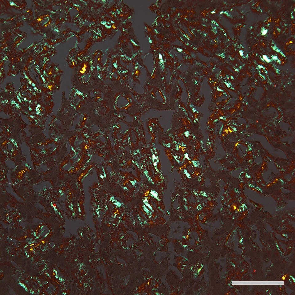
Original MICA image-reading exercise, unscored. No clinical narrative absent from the source is added.
- Describe visible findings before proposing an interpretation.
- Suggest an interpretation and at least one image limitation.
- Which clinical findings or investigations would complement this observation?
Reveal the explained reading
Amyloid deposits · interpretation and confirmation
- Tissue
- Liver
- Stain
- Congo red
- Reading
- Birefringence under polarised light
- Limitation
- The image does not type the protein
Morphological description
This Congo-red-stained section is viewed with polarised light; deposits are birefringent. Describe their distribution in the tissue. Colour alone does not type the protein.
Causes and diagnostic direction
AL, AA and ATTR have different mechanisms; micrograph colour cannot distinguish them.
Differential diagnoses
Staining may show optical pitfalls; interpret morphology, Congo red and polarisation together.
Pitfalls and limitations
Classic green is not the only possible colour; do not confuse birefringence with fluorescence.
Take-home message
Separate morphological findings, amyloid confirmation and identification of the deposited protein.
Sources
- Approach to the Diagnosis of Amyloidosis - PMC ↗
- Origins of a pervasive, erroneous idea: The “green birefringence” of Congo red‐stained amyloid - PMC ↗
Documentary review: 2026-10-12
Media source & rights
- Author
- Tulemo
- Original source
- Tulemo — Amyloid Liver Congo Red Bar=100um.jpg — CC BY-SA 4.0 — https://commons.wikimedia.org/wiki/File:Amyloid_Liver_Congo_Red_Bar%3D100um.jpg
- Licence
- CC BY-SA 4.0
- Changes
- Source rendition preserved. Full-size display converted to lossless WebP; thumbnail proportionally reduced to320px. No crop, medical retouching or alteration of the scale bar.
- Dimensions
- 960 × 960 px
- Check
- 2026-10-11