Original MICA image-reading exercise, unscored. No clinical narrative absent from the source is added.
- Describe visible findings before proposing an interpretation.
- Suggest an interpretation and at least one image limitation.
- Which clinical findings or investigations would complement this observation?
Reveal the explained reading
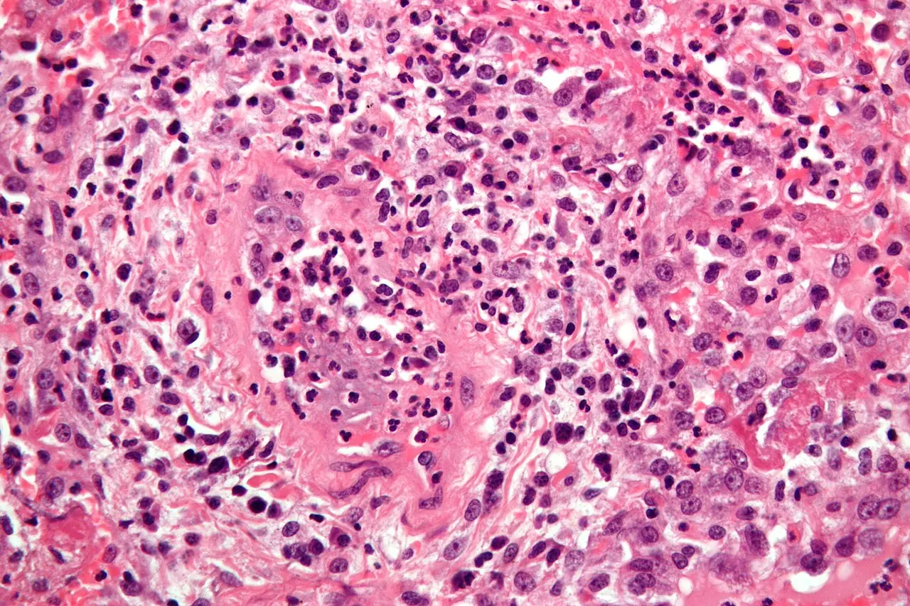
Granulomatosis with polyangiitis: micrograph
- Observation
- Inflammatory cells are visible in the selected field.
- Limitation
- Do not invent visible vasculitis in every field.
Morphological description
Inflammatory cells are visible in the selected field.
Source context
The source diagnosis is not established by this thumbnail alone.
Differential diagnoses
Consider other inflammation, infections and granulomas.
Pitfalls and limitations
Do not invent visible vasculitis in every field.
Take-home message
Describe structures actually present.
Sources
Documentary review: 2026-10-14
Media source & rights
- Author
- Nephron
- Original source
- Nephron · Granulomatose avec polyangéite : micrographie · CC BY-SA 3.0
- Licence
- CC BY-SA 3.0
- Changes
- Whole image retained; proportional resize and WebP conversion only. No clinical alteration. Derivative retains the original licence.
- Dimensions
- 1280 × 853 px
- Check
- 2026-10-14